Skin Cyst
Removal
Most skin cysts are benign — but a cyst that enlarges, inflames, or repeatedly gets infected deserves proper treatment, not another round of draining. The only way to prevent a cyst from coming back is to remove the entire sac wall. At Couture Dermatology and Laser, Dr. Chinonso Kagha Abisogun performs in-office surgical cyst excision with fine-line closure and a clear aftercare plan.
for low recurrence
anesthesia
Dermatologist
"Draining a cyst empties it. Excising the entire wall is what stops it from coming back."
A Sac Beneath
the Skin.
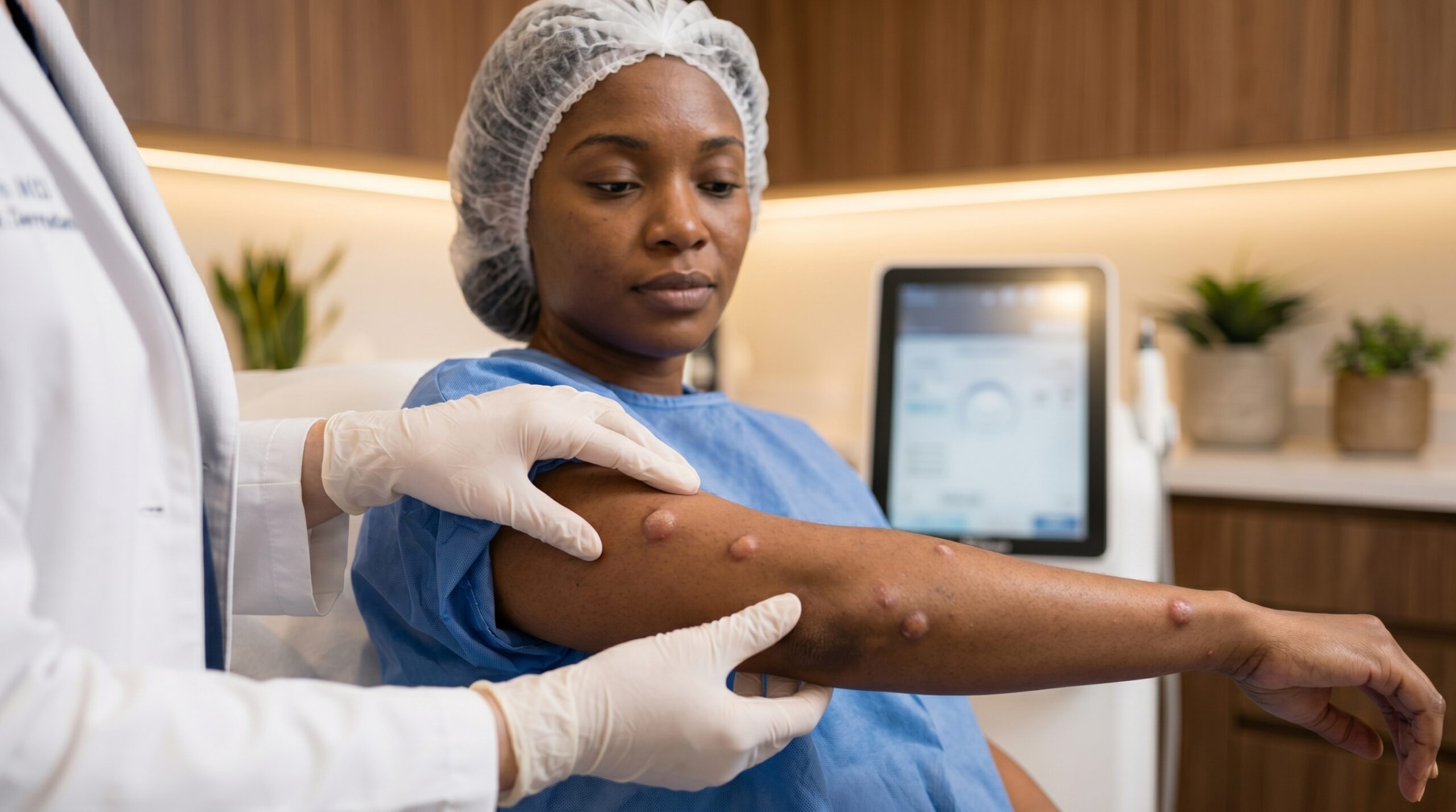
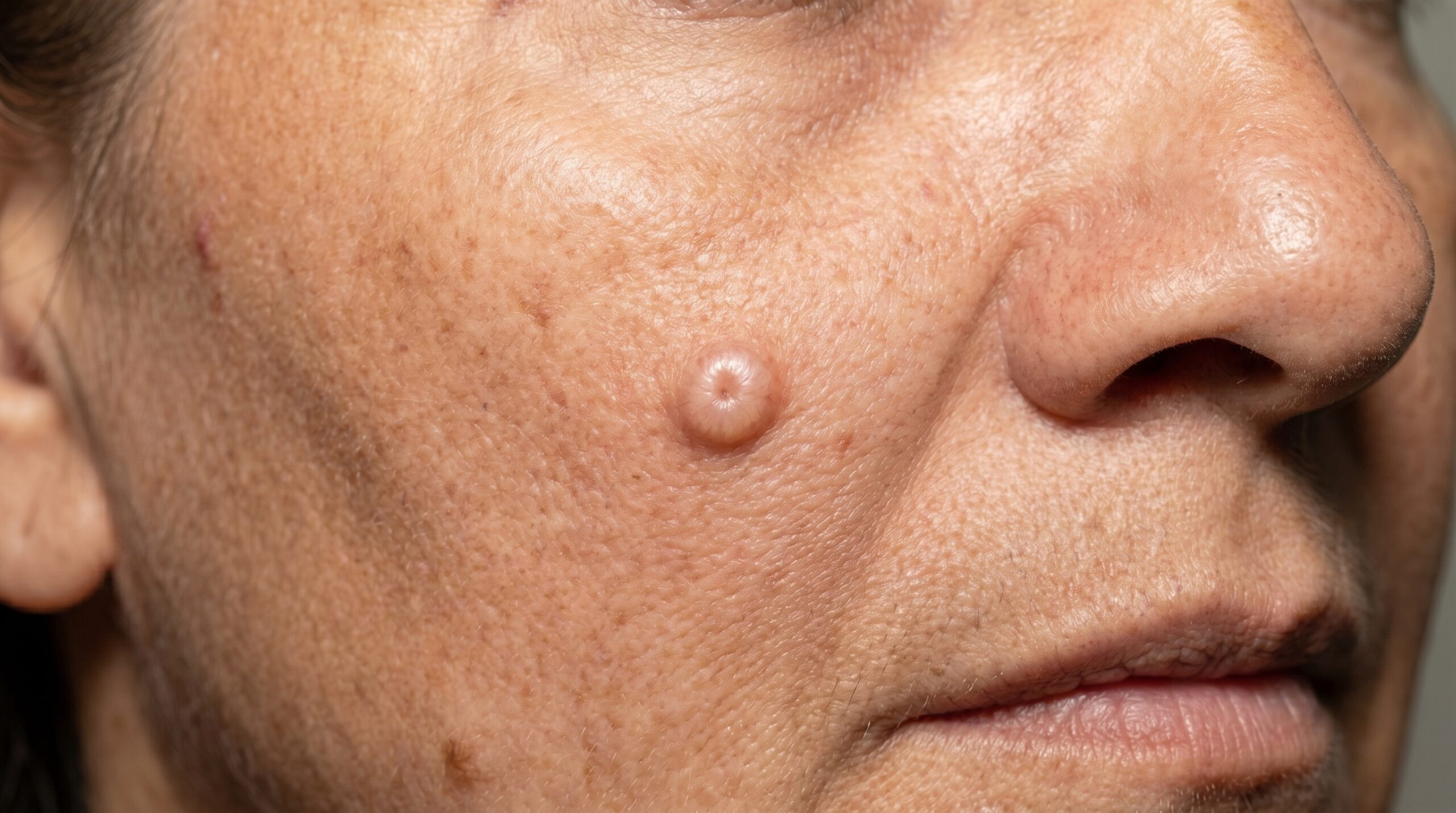
A skin cyst is a benign, enclosed sac lined by epithelium. The most common type is the epidermoid cyst — often called a "sebaceous cyst," though that is technically a misnomer. Epidermoid cysts are dome-shaped nodules, frequently with a small central punctum, filled with a soft keratin material. They appear most often on the face, neck, chest, and back.
Pilar (trichilemmal) cysts occur almost exclusively on the scalp, arise from the hair follicle root sheath, and tend to run in families. They feel firmer than epidermoid cysts and lack a punctum. Other types encountered in dermatology practice include milia (tiny keratin-filled cysts), digital mucous cysts near finger joints, and ganglion cysts.
Left alone, most cysts grow slowly and cause no symptoms. The problem arises when a cyst ruptures into the surrounding tissue, triggering an inflammatory reaction — the cyst becomes red, swollen, and tender, and may drain a cheesy or foul-smelling material. At that point, secondary bacterial infection is possible. Squeezing a cyst at home almost always makes this worse and increases the risk of scarring.
From Diagnosis
to Healed
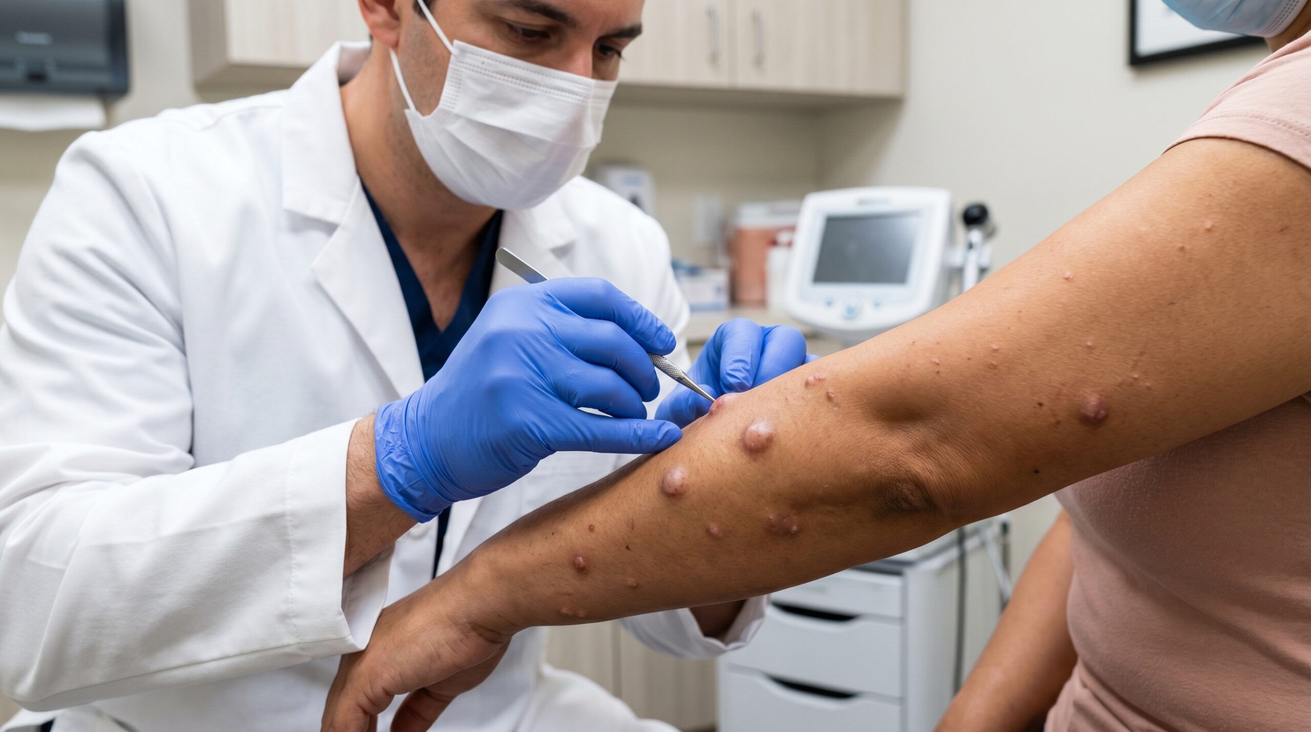
Cyst removal is a straightforward in-office procedure when the tissue is quiet. The process follows a deliberate sequence to ensure the cleanest possible excision and the best long-term outcome.
Clinical Evaluation
Dr. Chinonso examines the cyst, confirms benign characteristics, and determines whether it is currently suitable for excision or whether inflammation must be addressed first. Atypical features prompt a plan for pathology.
Diagnosis firstInflammation Control (if needed)
An acutely inflamed or infected cyst is not excised immediately. Incision and drainage, an intralesional corticosteroid injection, or a short antibiotic course settles the tissue before definitive surgery is scheduled.
4–6 weeks to quiet tissueSurgical Excision
Under local anesthesia, the cyst is excised through a small elliptical incision. The entire sac — wall and contents — is dissected out intact. Fragmentation of the wall increases recurrence risk, so care is taken to keep it whole.
20–45 minutesClosure & Healing
The wound is closed in layers with fine sutures to minimize the scar. Sutures are removed at 7 to 14 days. Scar-care instructions, including sun protection, are provided at the follow-up visit.
Sutures out in 1–2 weeksMatched to the
Clinical Picture
Not every cyst is treated the same way. The right procedure depends on whether the cyst is currently inflamed, its size, and its location. Here are the four approaches used at Couture Dermatology and Laser.
Definitive Treatment
Complete Surgical Excision
The gold-standard treatment for benign skin cysts. The entire cyst sac and its wall are removed intact through a small incision under local anesthesia. When the wall is fully excised, recurrence is rare. This is the approach used for stable, non-inflamed cysts.
Acutely Inflamed Cysts
Incision & Drainage
When a cyst is actively infected or severely inflamed, excision is deferred. A small incision is made to drain the contents and relieve pressure. This is a temporary measure — it resolves the acute episode but does not remove the cyst wall. Definitive excision is scheduled 4 to 6 weeks later.
Sterile Inflammation Without Infection
Intralesional Corticosteroid
For cysts that are inflamed but not frankly infected, a small intralesional injection of triamcinolone acetonide rapidly reduces swelling and tenderness within 48 to 72 hours. This settles the tissue so a clean excision can proceed electively without the risks that come with operating on acutely inflamed tissue.
Uncertain Lesions
Histopathological Review
Any excised tissue that looks atypical — rapidly enlarging, unusually firm, vascular, or with irregular features — is submitted to pathology. Benign cysts very rarely undergo malignant transformation, but changes in a long-standing lump warrant a histological diagnosis for peace of mind and clinical accuracy.
Clean Removal.
No Return.
Eliminates the Risk of Recurrence
When the entire cyst wall is excised intact, the epithelium that produces cyst contents is gone. Partial drainage, by contrast, leaves the sac in place and the cyst refills — often within weeks to months.
Prevents Repeated Infections
Cysts that have inflamed once tend to do so again. Repeated cycles of rupture and bacterial infection cause progressive fibrosis in the surrounding tissue, making each subsequent excision more technically demanding. Removing the cyst during a quiet window is the cleanest surgical outcome.
Fine-Line Scar Outcome
Careful elliptical incision design, intact sac removal, and layered closure produce a flat, well-healed linear scar that is typically far less conspicuous than a cyst that has been squeezed or repeatedly drained and has formed thick scar tissue of its own.
Accurate Diagnosis Confirmed
Excised tissue from any lesion with atypical features can be sent to pathology. For the vast majority of cysts, this confirms the benign diagnosis. For any lesion that turns out to be something unexpected, early detection changes outcomes significantly.
Quick Return to Normal Activity
Most patients experience mild soreness and localized swelling for a few days after excision and return to their regular routine within 24 to 48 hours. Sutures are removed at a short follow-up visit, typically 7 to 14 days later depending on location.
"A cyst that is drained but not excised has not been treated — it has been temporarily emptied."— Couture Dermatology and Laser
suture removal
Board-Certified
Every cyst removal is performed personally by Dr. Chinonso Kagha Abisogun, MD, FAAD — not delegated to support staff
Wall-Complete Excision
We prioritize removing the intact sac — not just draining contents — because half-measures lead directly to cyst recurrence
Staged When Appropriate
Inflamed cysts are treated first with I&D or intralesional steroid; excision is scheduled once tissue is quiet for the cleanest result
Pathology When Indicated
Atypical or rapidly changing lesions are sent for histopathological review — benign cysts rarely transform, but we verify what we remove
Common Cysts
Managed Here
Not all skin lumps are the same. Below are the cyst types most commonly evaluated and treated at Couture Dermatology and Laser. An accurate clinical diagnosis always comes first.
Epidermoid Cyst
The most common skin cyst. A dome-shaped nodule with a central punctum, lined by epidermis, filled with keratin. Often called a "sebaceous cyst," though that is not the correct term.
Pilar (Trichilemmal) Cyst
Found almost exclusively on the scalp, arising from the hair follicle root sheath. Tends to be firmer than epidermoid cysts, has no punctum, and often runs in families. Multiple cysts are common.
Inflamed or Infected Cyst
A cyst that has ruptured or become secondarily infected — red, tender, and swollen. Requires I&D or steroid injection first; excision follows once inflammation has fully resolved.
Milia
Small, superficial keratin cysts appearing as firm white or yellow papules, most often around the eyes and cheeks. Can be extracted under local anesthetic or removed with a fine lancet.
Digital Mucous Cyst
A myxoid cyst arising near finger joints or around the nail fold, often associated with underlying osteoarthritis. Treated by excision or intralesional injection depending on size and symptoms.
Recurrent Cyst
A cyst that has regrown after previous drainage or incomplete excision. Requires careful dissection through scar tissue to remove all residual sac wall — technically more demanding, but achievable.
Is Cyst Removal
Right for You?
- Anyone with a slowly enlarging, dome-shaped nodule they suspect is a skin cyst and want properly diagnosed and removed.
- Patients whose cyst has become inflamed, ruptured, or infected more than once and want a permanent solution, not repeated draining.
- Those with a cyst in a cosmetically sensitive location — face, neck, or décolletage — where a clean surgical scar is preferable to an enlarging lump.
- Patients with multiple scalp pilar cysts causing discomfort or hair-loss pressure at the cyst site.
- Anyone with a previously "drained" cyst that has refilled and wants it properly excised this time, including dissection through any existing scar tissue.
- Patients with a changing lump — rapid growth, hardening, or irregular borders — who need a clinical assessment and, if warranted, pathological confirmation.
What to Expect Honestly
Cyst excision does leave a scar — any surgical incision does. The goal is a flat, fine-line scar that fades over 6 to 12 months and is far less conspicuous than the cyst itself, particularly for cysts that have been inflamed and have already distorted the overlying skin.
Patients with a personal or family history of keloid or hypertrophic scarring should discuss this with Dr. Chinonso before proceeding. The risk of recurrence after complete wall excision is low, but no surgical excision carries a zero recurrence rate — particularly for cysts that required dissection through dense scar tissue from prior procedures.
Other Skin Growths
We Address
Patients presenting with cysts often have other benign skin growths that can be evaluated and treated at the same visit. These are the procedures most commonly combined with cyst removal.
Skin Tag Removal
Small, soft, pedunculated growths occurring at friction sites — neck, axillae, groin. Removed quickly in the same visit using snip excision or electrosurgery with no sutures required.
Explore skin tag removal →Mole Removal
Benign moles that are irritated, enlarging, or cosmetically bothersome can be shave-excised or surgically removed. Any mole with atypical features is sent for pathological review as standard practice.
Explore mole removal →Keloid Treatment
Patients prone to keloid or hypertrophic scarring benefit from a pre-surgical discussion and a post-excision management plan — including intralesional steroid injections and silicone gel — to minimize abnormal scar formation at the excision site.
Explore keloid treatment →Skin Rash Evaluation
Cysts can be mistaken for other dermatological conditions, and vice versa. If the diagnosis is uncertain — or if there are coexisting skin changes — a thorough medical dermatology evaluation ensures nothing is missed.
Explore medical dermatology →Results That Speak for Themselves
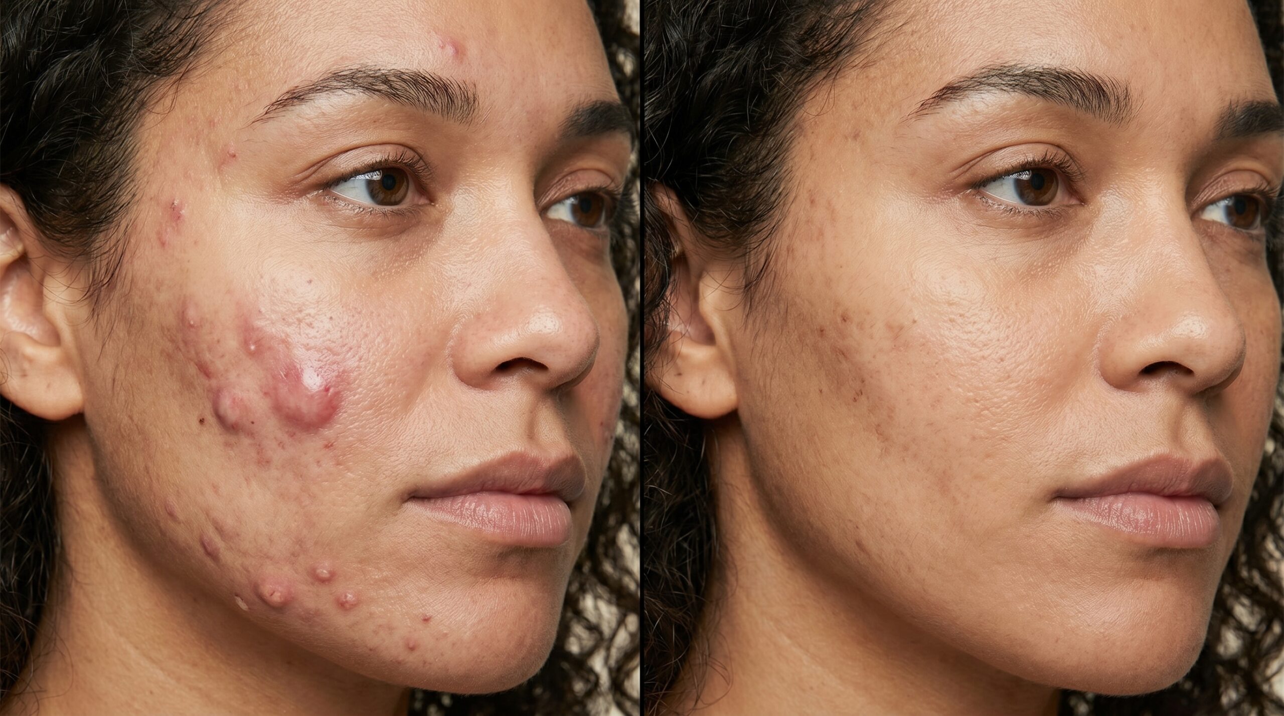
Individual results vary. Final scar appearance continues to improve for 6 to 12 months after excision with appropriate scar care and sun protection.
Remove It Once.
Remove It Completely.
A cyst that keeps coming back has not been fully treated. At Couture Dermatology and Laser, Dr. Chinonso evaluates every lesion clinically, determines the right timing for excision, and performs complete wall removal with careful closure. No shortcuts. No repeat visits for the same cyst.
Sat · By Appointment Only
"I had the same cyst drained twice at urgent care and it came back both times. Dr. Chinonso explained exactly why — they never removed the wall — and then excised it properly. It has been six months and there is nothing there. The scar is barely visible."
Marcus T.
Verified Patient · Beverly Hills
Frequently
Asked Questions
Direct answers to the questions patients most commonly ask before their cyst removal consultation — on types, timing, recurrence, and what the procedure actually involves.
The terms are often used interchangeably by patients, but clinically they are different. An epidermoid cyst is lined by normal epidermis and filled with keratin — it has a visible central punctum and is the most common type on the face, neck, and trunk. A true sebaceous cyst arises from the sebaceous gland and is actually less common. Most lumps patients call "sebaceous cysts" are in fact epidermoid cysts. Pilar cysts on the scalp originate from hair follicle root sheaths and do not have a punctum.
Not every cyst requires removal. Small, stable, asymptomatic cysts can simply be monitored. However, removal is advisable if the cyst is enlarging, has become inflamed or infected, is painful, is located somewhere that causes functional or cosmetic concern, or if the diagnosis is uncertain and pathology is needed. Recurrently inflamed cysts are best excised during a quiet period to prevent future episodes.
Draining a cyst — squeezing it at home or having it incised without full removal — empties the contents but leaves the cyst wall (sac) intact in the dermis. Because the sac is lined by epithelium that continues producing keratin or sebaceous material, the cyst simply refills — often within weeks to months. The only definitive treatment that prevents recurrence is complete surgical excision of the entire cyst wall.
No. Excising an acutely infected or severely inflamed cyst is technically difficult and significantly raises the risk of incomplete removal and recurrence, because the tissue planes are distorted and fragile. The standard approach is to perform incision and drainage first — or in some cases an intralesional corticosteroid injection for inflamed but not frankly infected cysts — and then schedule a clean excision 4 to 6 weeks later once the inflammation has fully resolved.
The area is numbed with a local anesthetic injection, so the procedure itself is not painful. The cyst is excised through a small elliptical or linear incision, the sac is carefully dissected out intact, and the wound is closed with sutures. Most procedures take 20 to 45 minutes depending on size and location. Sutures are removed at 7 to 14 days. There is typically mild soreness and swelling for a few days; most patients return to normal activities within 24 to 48 hours.
Yes — any surgical excision leaves a scar, but the goal is a fine, flat, well-healed linear scar that is far less conspicuous than the cyst itself. Dr. Chinonso closes each wound with careful layered sutures and provides scar-care instructions including sun protection. Patients who are prone to hypertrophic or keloidal scarring will have their individual risk discussed before the procedure.