Melanoma
Detection & Care
Melanoma is the most serious form of skin cancer — not because it is the most common, but because it can spread. Unlike basal cell and squamous cell carcinomas, melanoma has genuine metastatic potential. Caught early, at a thin and localized stage, it is highly treatable. That window is exactly what a trained dermatologist is positioned to protect.
criteria
when caught early
Dermatologist
"A changing lesion is not something to watch at home. It is something to have examined today."
Melanocytes.
Moles. Margins.
Melanoma originates in melanocytes — the cells responsible for pigment production. It may arise within an existing mole or appear as an entirely new lesion on skin that previously looked unremarkable. UV radiation is the leading environmental risk factor, though melanoma also occurs on sun-protected sites, which is part of why comprehensive skin examination matters.
Diagnosis requires biopsy. Clinical appearance, even with dermoscopy, cannot definitively confirm melanoma — histopathologic evaluation by a dermatopathologist does. The biopsy specimen determines the Breslow depth (how thick the tumor is in millimeters), ulceration status, and mitotic rate. These three factors drive staging and, critically, every subsequent treatment decision.
Early-stage melanoma — thin, localized, without nodal involvement — is treated definitively by wide local excision. It is at the later stages, when melanoma has reached lymph nodes or distant organs, that management becomes significantly more complex. This is why the dermatologist's first responsibility is to find it before it progresses.
From Detection
to Definitive Care
Melanoma management follows a precise sequence. Each step informs the next, and no treatment decision is made before the pathology is known.
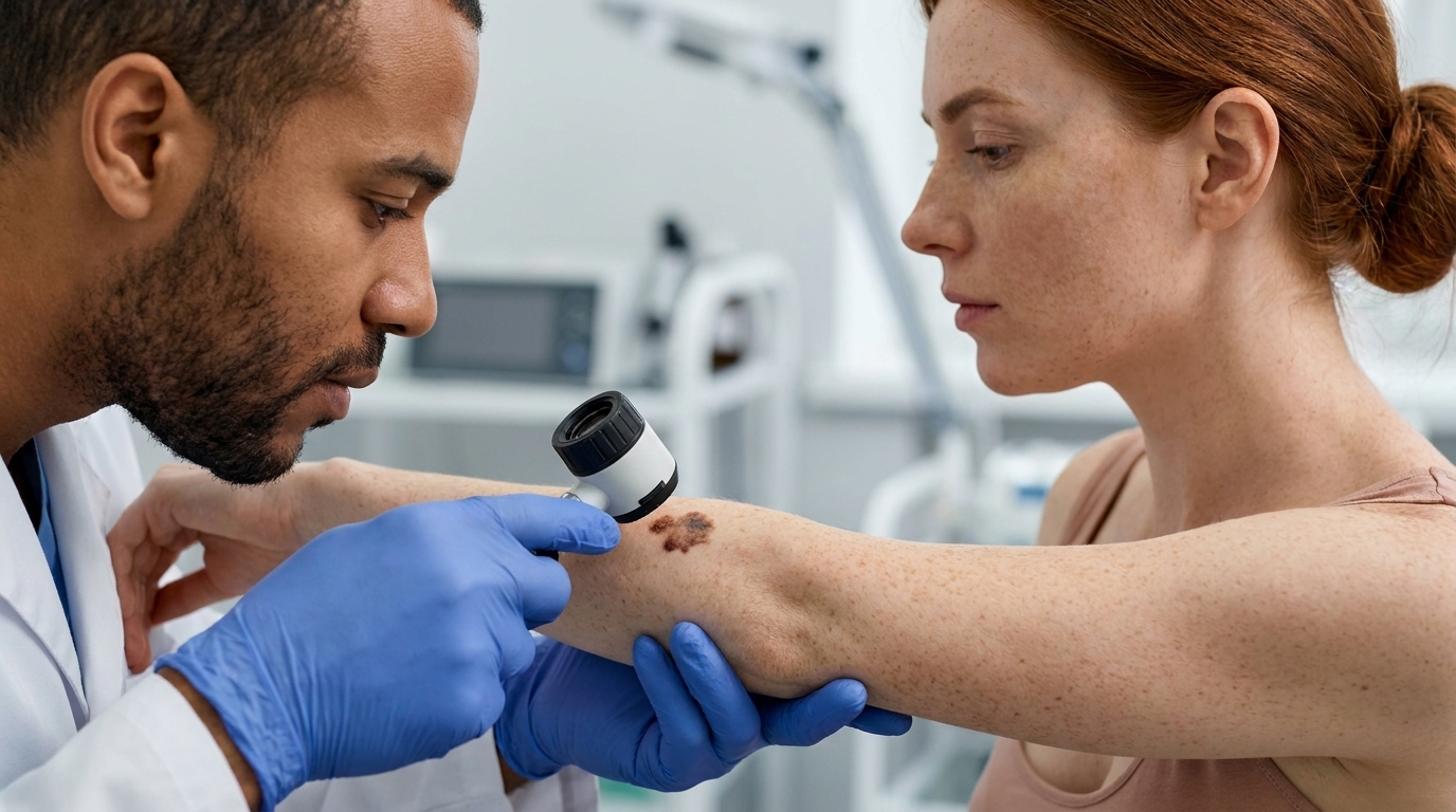
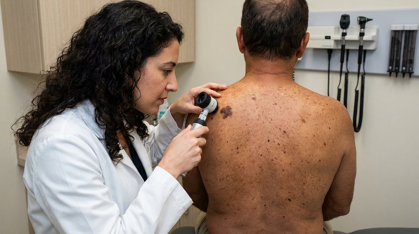
Full-Body Skin Exam
A thorough head-to-toe examination, including dermoscopy, to identify suspicious lesions that meet ABCDE criteria or appear as an "ugly duckling" among other moles.
Includes dermoscopyExcisional Biopsy
Suspicious lesions are removed with a narrow margin and sent to dermatopathology. The report provides diagnosis, Breslow depth, ulceration, and mitotic rate — the data that drives every subsequent decision.
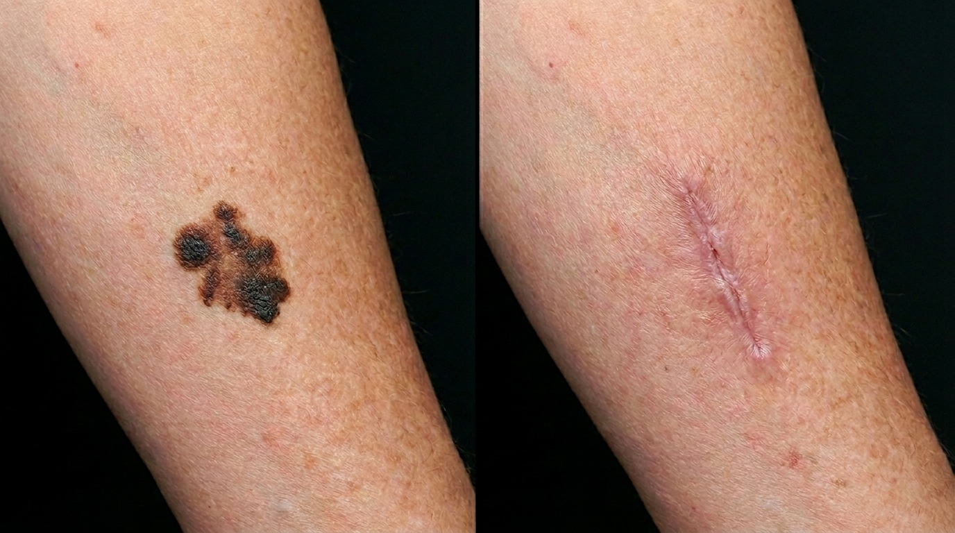
Same or next visitWide Local Excision
Confirmed melanoma requires re-excision with wider margins determined by Breslow depth — from 0.5 cm for in-situ disease to 2 cm for tumors thicker than 2 mm — following current NCCN guidelines.
Margin guided by depthSurveillance & Coordination
After definitive excision, a structured follow-up schedule is established. Higher-stage cases are coordinated with surgical oncology for sentinel lymph node biopsy and, where indicated, medical oncology for systemic therapy.
Lifelong monitoringHow Melanoma Is
Found and Managed
The clinical tools used at each stage of melanoma care serve distinct purposes. Knowing when to use each one is the difference between early diagnosis and a missed window.
Detection
Dermoscopy
A handheld optical device that illuminates and magnifies skin structures below the surface, revealing pigment network patterns, vascular architecture, and regression features that cannot be evaluated by the naked eye. Dermoscopy significantly improves diagnostic accuracy for melanocytic lesions and reduces unnecessary biopsies of benign moles.
Diagnosis
Excisional Biopsy
The only way to definitively diagnose melanoma. The entire lesion is removed with a 1–3 mm margin, preserved in formalin, and evaluated by a dermatopathologist. The resulting report specifies diagnosis, Breslow depth, Clark level, ulceration, and mitotic rate — the complete staging dataset for treatment planning.
Treatment
Wide Local Excision
Surgical removal of the confirmed melanoma site with guideline-specified margins. For melanoma in situ, 0.5–1 cm margins are standard. Invasive melanoma requires 1–2 cm margins based on Breslow depth. WLE is the definitive treatment for localized early-stage disease and is curative in the majority of thin melanomas.
High-Risk & Advanced
Multidisciplinary Care
Melanomas with a Breslow depth above 0.8 mm, ulceration, or high mitotic rate require discussion of sentinel lymph node biopsy for accurate nodal staging. Metastatic melanoma is managed with immunotherapy (checkpoint inhibitors) and targeted therapy (BRAF/MEK inhibitors), coordinated with medical and surgical oncology.
The Case for
Acting Now.
Stage Determines Prognosis
A thin melanoma diagnosed at Stage IA carries an excellent long-term prognosis and is treated with surgery alone. The same tumor, found two years later at Stage III with nodal involvement, requires a fundamentally different — and far more difficult — course of treatment. Stage at diagnosis is the single most important prognostic variable.
Dermoscopy Catches What Eyes Miss
Studies consistently show that dermoscopy improves melanoma detection sensitivity by 10–27% over unaided visual inspection alone. Subtle early features — regression structures, atypical vascular patterns, irregular pigment networks — become visible only under magnified illumination. This is not a luxury tool; it is standard of care in experienced hands.
Breslow Depth Guides Every Decision
The thickness of the primary tumor in millimeters — the Breslow depth — determines excision margins, informs the sentinel lymph node biopsy decision, drives staging, and predicts recurrence risk. This single measurement is the keystone of the entire melanoma treatment plan, which is why biopsy technique matters.
Acral Melanoma in Darker Skin Tones
Acral lentiginous melanoma — the subtype occurring on palms, soles, and beneath the nails — is the predominant form in patients with darker skin, where it is frequently diagnosed at a more advanced stage because it is not in a sun-exposed location and may not be on the standard self-exam checklist. Total-body skin examination includes these sites.
Surveillance Prevents Second Primaries
A personal history of melanoma raises the risk of a second primary melanoma by approximately 5–10 times compared to the general population. Structured surveillance — typically every 3–6 months in the first two years and annually thereafter — combined with total-body photography provides the best framework for catching recurrence or new primaries at the earliest possible stage.
"A melanoma found at 0.5 mm is a surgical problem. Found at 3 mm with nodal involvement, it becomes a systemic one. The exam that changes the equation takes twenty minutes."— Couture Dermatology and Laser
exams recommended
Board-Certified
Every skin examination is performed personally by Dr. Chinonso Kagha Abisogun, MD, FAAD — Fellow of the American Academy of Dermatology
Dermoscopic Evaluation
Dermoscopy at every full-body exam — magnified subsurface evaluation of pigmented lesions that visual inspection alone cannot provide
Guideline-Driven Excision
Wide local excision margins based on current NCCN guidelines, with Breslow depth as the determining variable — not estimate or convention
Coordinated Care
Advanced or metastatic disease is coordinated with surgical and medical oncology — the dermatologist does not manage these cases in isolation
Six Subtypes.
One Disease.
Melanoma is not a single clinical entity. Recognizing the distinct presentations of each subtype is essential to catching the ones that do not fit the classic picture.
Superficial Spreading
The most common subtype. Grows radially along the skin surface before invading deeper. Often presents as an asymmetric, multi-colored flat lesion on the trunk or extremities.
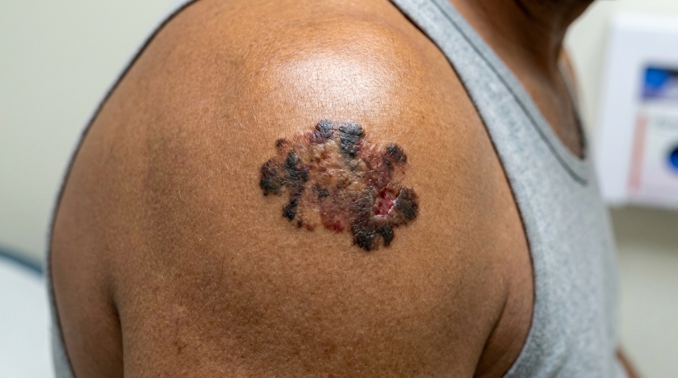
Nodular Melanoma
Grows vertically and aggressively from the outset, often presenting as a rapidly enlarging, dome-shaped, dark nodule. May bleed or ulcerate. Breslow depth at diagnosis is frequently high.
Lentigo Maligna
A slow-growing subtype arising on chronically sun-damaged skin — typically the face and neck in older patients. Appears as a flat, irregularly pigmented tan or brown patch; may evolve to invasive lentigo maligna melanoma over years.
Acral Lentiginous
Occurs on the palms, soles, and beneath the nails — not sun-exposed sites. The most common melanoma subtype in patients with darker skin tones. Frequently diagnosed at a later stage due to its non-typical location.
Amelanotic Melanoma
Lacks visible pigment, presenting as a pink, red, or skin-colored lesion. Easily mistaken for a cyst, pyogenic granuloma, or scar. Dermoscopy reveals vascular patterns that raise suspicion even without pigmentation.
Desmoplastic Melanoma
A rare, spindle-cell variant, often amelanotic, that arises on the head and neck. Associated with high local recurrence after excision. Diagnosis requires careful histopathology; wide margins are essential.
When Should
You Come In?
- Anyone who has noticed a mole or spot that has changed in size, shape, color, or texture — or started to bleed, crust, or itch.
- Patients with a personal or family history of melanoma, who require more frequent surveillance examinations than the general population.
- Those with a large number of moles, a history of dysplastic (atypical) nevi, or prior biopsy-confirmed dysplastic nevi with severe atypia.
- Individuals with significant UV exposure history — including indoor tanning, frequent sunburns, or occupational sun exposure — particularly if it occurred in youth.
- Patients with darker skin tones who want proper evaluation of lesions on the palms, soles, or beneath the nails — sites where melanoma is not related to UV exposure.
- Anyone who has not had a full-body skin examination in the past year and is over 40, immunocompromised, or has any of the above risk factors.
A Direct Conversation About Prognosis
Melanoma is serious, and we will not minimize that. What is equally true is that the majority of melanomas diagnosed in a dermatology office — because patients came in promptly — are thin, early-stage, and surgically curable. The prognosis for Stage I melanoma is excellent. Prognosis for Stage IV is substantially harder.
The goal of a skin examination is not to find something frightening — it is to find something early enough that the treatment is straightforward. Dr. Chinonso will discuss any finding directly, explain the pathology report in plain terms, and make certain you understand your options before any next step is taken.
Melanoma in Context
of Skin Cancer Care
Melanoma rarely exists in isolation. Patients at risk for melanoma often carry risk factors for other skin cancers as well. These related services complete the picture.
Mole Evaluation & Mapping
For patients with many moles or a history of atypical nevi, total-body photography provides a documented baseline. Interval comparisons make subtle changes visible that would otherwise go unnoticed between annual visits.
Explore mole evaluation →Basal Cell Carcinoma
The most common skin cancer. Rarely metastasizes but can cause significant local tissue destruction if neglected. Patients with a history of BCC have an elevated lifetime risk of both BCC recurrence and melanoma.
Explore BCC treatment →Squamous Cell Carcinoma
The second most common skin cancer. SCC can metastasize, particularly in high-risk locations and in immunocompromised patients. Often arises from actinic keratoses, making precancer management a meaningful prevention strategy.
Explore SCC treatment →Actinic Keratosis
Rough, scaly precancerous patches caused by cumulative UV damage. While actinic keratoses do not become melanoma, they signal significant sun damage — and patients with many AKs should also be examined closely for melanocytic changes.
Explore AK treatment →From Diagnosis to Clear Margins
Clinical outcomes vary based on melanoma subtype, Breslow depth, stage at diagnosis, and individual patient factors. Photography shown for educational purposes. Images represent individual clinical cases.
Early Detection.
Every Exam Counts.
The most valuable thing a dermatologist does for a patient at risk for melanoma is perform a thorough skin examination before anything has changed enough to be obvious. At Couture Dermatology and Laser, Dr. Chinonso examines every patient personally, documents findings carefully, and acts on anything suspicious the same day. No lesion is dismissed without dermoscopic evaluation.
Sat · By Appointment Only
"I came in for what I thought was just a routine mole check. Dr. Chinonso found a lesion on my back I had no idea about, biopsied it the same visit, and called me personally with the results. It was early-stage melanoma. The excision was two weeks later. I cannot overstate how important that appointment was."
James T.
Verified Patient · Beverly Hills
Frequently
Asked Questions
Honest answers to the questions patients ask most often before a melanoma evaluation — on warning signs, diagnosis, surgical margins, risk factors, and surveillance.
Melanoma arises from melanocytes — the pigment-producing cells of the skin. Unlike basal cell and squamous cell carcinomas, which rarely spread beyond the skin, melanoma has significant metastatic potential: it can travel through lymphatic and blood vessels to distant organs. This is why early detection is so consequential. Caught at a thin, early stage, the prognosis is excellent; advanced-stage disease is far more difficult to treat.
The ABCDE criteria are the most reliable self-exam guide: Asymmetry (one half differs from the other), Border irregularity (jagged, notched, or blurred edges), Color variation (multiple shades of brown, black, red, white, or blue in one lesion), Diameter greater than 6mm, and Evolving (any lesion that is changing in size, shape, color, or beginning to bleed or crust). The "ugly duckling" sign is equally useful — any lesion that simply looks different from your other moles deserves evaluation.
Dr. Abisogun performs a comprehensive total-body skin examination and uses dermoscopy — a handheld device that magnifies and illuminates subsurface skin structures — to evaluate pigmented lesions that cannot be assessed with the naked eye alone. Suspicious lesions are biopsied the same day or at a closely scheduled follow-up. The biopsy specimen is sent to a dermatopathologist for histopathologic diagnosis including Breslow depth measurement.
The primary treatment for localized melanoma is wide local excision — surgical removal of the tumor with a margin of normal surrounding skin. Margin width (0.5 cm to 2 cm) is determined by the Breslow depth of the lesion per NCCN guidelines. For melanomas with a Breslow depth above 0.8 mm, sentinel lymph node biopsy is discussed for accurate staging. Advanced or metastatic disease requires coordination with surgical and medical oncology, where immunotherapy and targeted (BRAF/MEK inhibitor) therapy have transformed outcomes.
Risk factors include a personal or family history of melanoma, a large number of moles (particularly atypical or dysplastic nevi), fair skin that burns easily, a history of blistering sunburns — especially in childhood — prior use of tanning beds, and immunosuppression. Melanoma also occurs in people of all skin tones: acral lentiginous melanoma, which appears on the palms, soles, and under the nails, is the most common subtype in darker-skinned individuals and is often diagnosed later because it is not in a sun-exposed site.
Annual full-body skin examinations are recommended for most adults. Patients with a personal history of melanoma, multiple atypical nevi, or a first-degree relative with melanoma should be seen every 3 to 6 months. Between visits, monthly self-examination using the ABCDE criteria is an important early-warning practice. Any lesion that changes, bleeds, or simply catches your attention warrants a prompt appointment — do not wait for the annual exam.