Mole Removal
& Evaluation
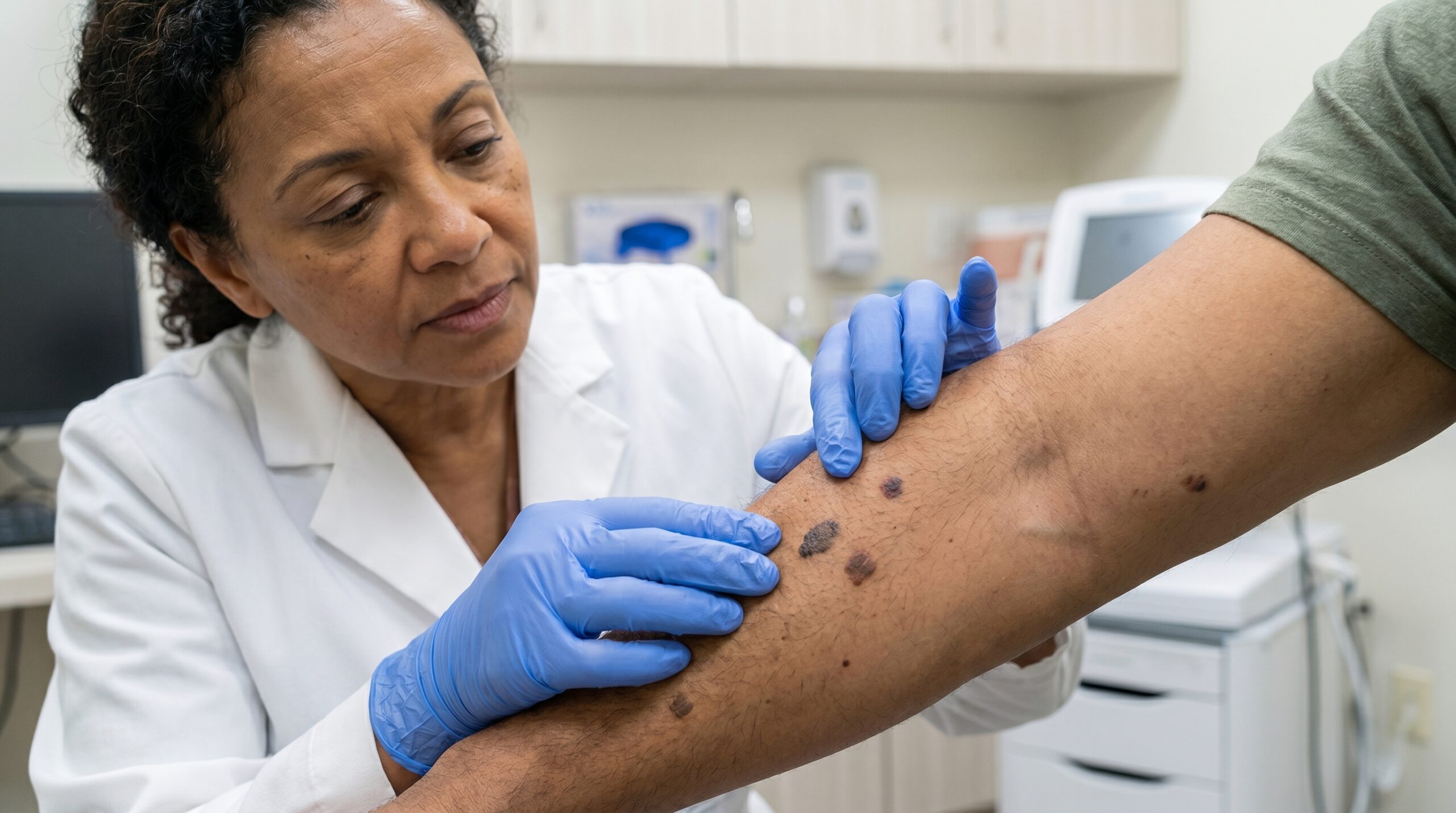
Most moles are harmless — but some change, and a small number can develop into melanoma. At Couture Dermatology and Laser, every mole evaluation starts with dermoscopy and a trained clinical eye. Whether you need a full-skin examination, monitoring of atypical nevi, or the removal of a mole that's changing or simply uncomfortable, the approach is methodical, thorough, and backed by pathology when it counts.
mole evaluation
to pathology always
Dermatologist
"A suspicious mole is never lasered off — the tissue it holds is what makes a diagnosis possible."
When to Watch.
When to Act.
Moles — clinically called melanocytic nevi — are benign clusters of pigment-producing cells that most people develop between childhood and their mid-30s. The vast majority are stable and harmless across a lifetime. The clinical question is always whether a particular mole belongs in that majority.
Dermoscopy — examining a mole with a polarized handheld lens — reveals structural patterns invisible to the naked eye: irregular pigment networks, atypical vascular patterns, and regression structures that suggest a lesion warrants closer scrutiny or removal. At Couture Dermatology and Laser, dermoscopy is standard at every mole evaluation.
The dual responsibility of mole care is straightforward: monitor what is stable, and promptly evaluate what changes. Any mole that bleeds, itches, grows, or simply looks different from your others — the "ugly-duckling" sign — earns a same-appointment evaluation, not a "wait and see."
From First Look
to Clear Answer
Mole evaluation at Couture Dermatology is not a rushed glance. Every step — from full-skin exam to pathology review — is designed to give you a definitive clinical answer.
Full-Skin Examination
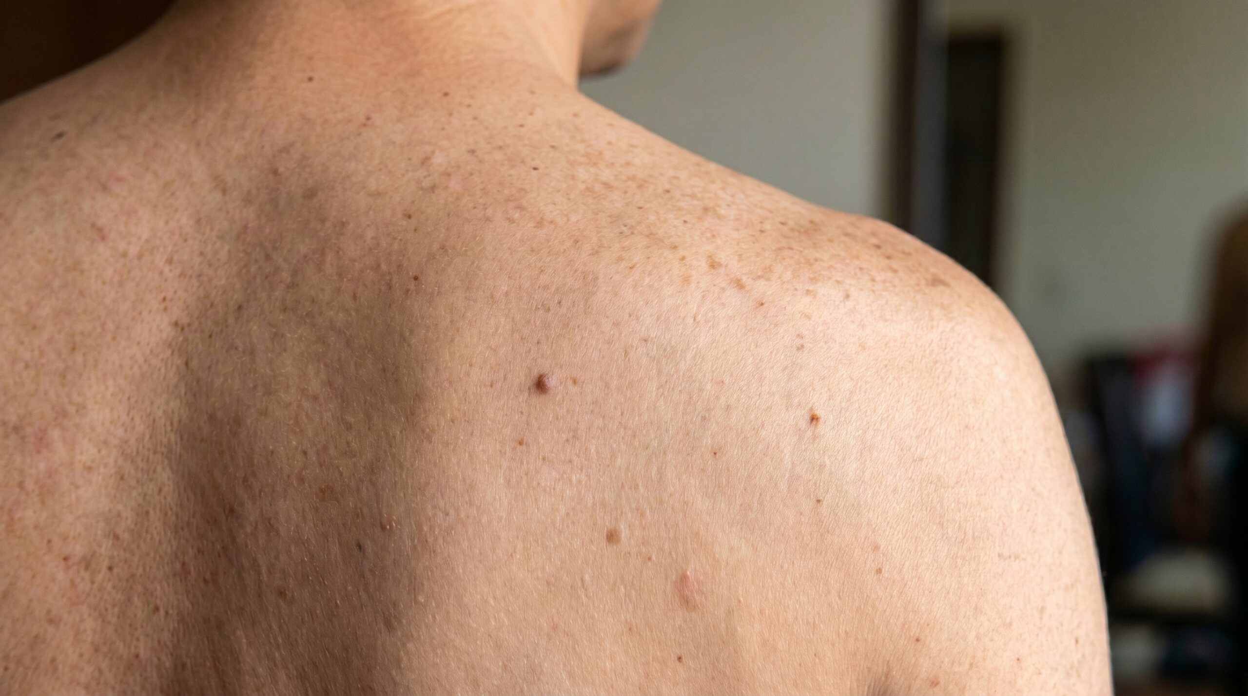
A head-to-toe skin check identifies all existing nevi, documents their characteristics, and flags any lesion that warrants dermoscopic assessment or immediate removal.
Dermoscopy includedClinical Decision
Each mole is classified: monitor, refer for monitoring, or remove. The decision is driven by clinical and dermoscopic criteria — not patient anxiety or cosmetic preference alone.
ABCDE + ugly-ducklingRemoval Procedure
Raised benign moles are removed by shave technique under local anesthesia. Any flat or clinically concerning lesion is excised with a surgical margin. Both are same-appointment procedures.
15–30 minutesPathology & Follow-up
All removed specimens with any clinical concern are sent for histological review. Results are communicated promptly, with further treatment recommended if margins or diagnosis require it.
Results in 1–2 weeksThe Right Tool
for Each Mole
Technique selection follows clinical logic. A cosmetically bothersome raised nevus is handled differently from a flat lesion with irregular dermoscopic features — and both are different from a monitoring case.
Raised Benign Moles
Shave Removal
A thin blade shaves the mole flush with or slightly below the skin surface under local anesthesia. This leaves a small, flat pink mark that fades over weeks to months. Ideal for benign raised nevi that rub on clothing or are cosmetically bothersome — not appropriate for flat or clinically atypical lesions.
Flat or Atypical Lesions
Surgical Excision
Full-thickness removal with a scalpel, closed with sutures. Used for flat moles, any lesion with clinical or dermoscopic features of concern, and congenital nevi requiring complete removal. Margins can be assessed histologically — which is why suspicious moles are always excised, never lasered or burned.
Structural Assessment
Dermoscopy
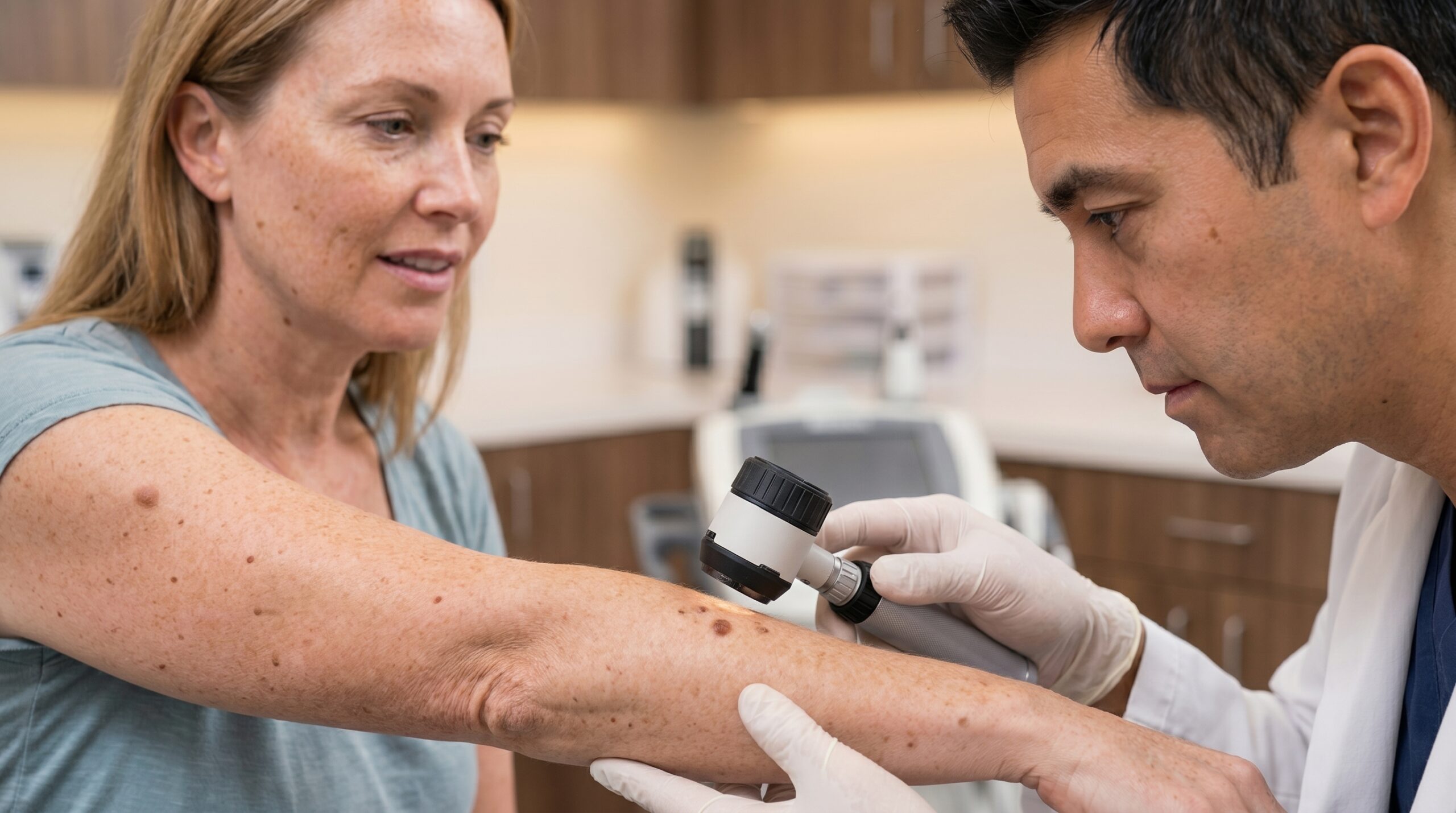
A polarized handheld lens eliminates surface glare and reveals subsurface pigment architecture, vascular patterns, and regression features invisible to the naked eye. Dermoscopy significantly improves diagnostic accuracy for distinguishing benign nevi from early melanoma compared to visual inspection alone.
Higher-Risk Patients
Monitoring & Serial Photography
Patients with multiple atypical nevi, a personal or family history of melanoma, or large congenital nevi benefit from structured surveillance with serial dermoscopic photography to detect subtle change over time — the kind of change that tells a clinician when a watchful mole has crossed a threshold.
Clear Skin.
Confirmed Results.
Early Detection When It Matters Most
Melanoma caught at Stage I has a five-year survival rate above 98%. A routine mole check that identifies an early-stage lesion is not a cosmetic appointment — it is a potentially life-altering one. Dermoscopy improves detection sensitivity well beyond the naked eye.
Diagnosis Confirmed by Pathology
When a mole is removed because it looks suspicious, the tissue goes to a dermatopathologist. You receive a histological diagnosis, not a clinical guess. If margins are incomplete or the pathology reveals dysplasia, a clear next step is available — none of which is possible if the tissue was destroyed by laser.
Cosmetic Removal Done Cleanly
Moles that catch on clothing, sit in awkward locations, or have simply bothered a patient for years can be removed efficiently under local anesthesia in a single appointment. The resulting mark is small and fades with time — though patients should understand that any removal leaves some trace.
Peace of Mind, Grounded in Evidence
A "this looks fine" from a board-certified dermatologist who examined the lesion with dermoscopy carries a very different weight than reassurance based on a surface glance. Patients leave with a genuine clinical assessment — and a clear plan if anything needs follow-up.
A Structured Surveillance Plan for High-Risk Patients
Multiple atypical moles, a family history of melanoma, or prior dysplastic nevi are not reasons for anxiety — they are reasons for a clear monitoring protocol. Serial photography and defined examination intervals convert risk into a manageable routine.
"The mole that looks fine today is the one worth documenting — so we know exactly what 'changed' means six months from now."— Couture Dermatology and Laser
results when needed
Board-Certified
Every mole evaluation is performed personally by Dr. Chinonso Kagha Abisogun, MD, FAAD — a Fellow of the American Academy of Dermatology
Dermoscopy Standard
No evaluation relies on the naked eye alone — polarized dermoscopy is used at every mole check to assess subsurface structure
Pathology Always
Clinically concerning moles are excised and sent to a dermatopathologist — suspicious lesions are never lasered or destroyed without diagnosis
Clear Communication
Patients receive a plain-language explanation of their findings, their options, and any follow-up required — never vague reassurance
Not All Nevi
Are the Same
The word "mole" covers a wide range of lesions with different risk profiles, appearances, and management strategies. Accurate classification is the starting point for appropriate care.
Common Acquired Nevi
The standard brown moles that develop after birth, typically uniform in color and well-defined. Usually benign and require only monitoring — removal is elective.
Congenital Nevi
Moles present at birth, ranging from small to large. Larger congenital nevi carry an elevated lifetime risk and may warrant surveillance or prophylactic removal depending on size and features.
Atypical / Dysplastic Nevi
Moles with irregular borders, uneven pigmentation, or diameters over 6mm. Histologically, dysplastic nevi show abnormal cell architecture. Those with high-grade dysplasia typically require excision with clear margins.
Blue Nevi
Blue or gray-blue lesions caused by melanocytes deep in the dermis. Most are benign, but cellular blue nevi or those that change warrant biopsy to rule out blue nevus-associated melanoma.
Spitz Nevi
Pink or reddish dome-shaped lesions often seen in children and young adults. Dermoscopic features can overlap with early melanoma, making biopsy the safe standard for any Spitz-pattern lesion.
Warning Signs (ABCDE)
Any mole with asymmetry, irregular borders, multiple colors, a diameter over 6mm, or recent change should be evaluated promptly. The ugly-duckling sign — a mole that simply looks different from your others — is an equally valid reason to seek an appointment.
Do You Need
a Mole Check?
- Anyone with a mole that has changed in size, shape, or color in recent weeks or months — change is the most clinically significant warning sign.
- Patients with a mole that bleeds without injury, itches persistently, or has developed an irregular, notched border.
- Those with more than 50 moles, a personal history of atypical nevi, or a first-degree relative who has had melanoma.
- Adults who have never had a comprehensive full-skin examination by a dermatologist, particularly those with fair skin or significant lifetime sun exposure.
- Patients with a raised or uncomfortable mole that catches on clothing, jewelry, or seatbelts and who want clean, same-visit removal.
- Anyone who has simply noticed a mole they don't remember having before — or one that looks different from the rest. A quick evaluation is always the right call.
What the Evaluation Will Tell You
A mole check is not a pass-or-fail test. The outcome is clinical clarity: a determination of whether a lesion is stable and can be monitored, warrants removal for diagnostic or cosmetic reasons, or needs excision and pathology because of features that don't fit the benign pattern.
Cosmetic removal is a legitimate reason to have a mole removed — but patients should understand that any technique, including shave removal, leaves a mark. The goal is to trade a raised or pigmented mole for a small, flat, fading scar. Dr. Chinonso will be direct about expected outcomes for your specific lesion before any procedure is performed.
Address the
Full Picture
A mole evaluation often surfaces related concerns — from other skin lesions that need attention to a broader skin-cancer surveillance plan. These services complete that picture.
Melanoma
When a mole removal returns a melanoma diagnosis, the clinical pathway continues: re-excision for clear margins, staging, and coordination with oncology if indicated. Early-stage melanoma found during a routine check is highly treatable.
Explore melanoma care →Skin Tags
Soft, flesh-colored growths that patients sometimes confuse with moles. Skin tags are benign fibrovascular lesions — not nevi — and are removed quickly by snip excision or cautery in the same visit as a mole evaluation.
Explore skin tag removal →Basal Cell Carcinoma
The most common skin cancer, often found during the same full-skin exam as a mole check. A pearly, pink, or ulcerated lesion that doesn't heal is the classic presentation — and a full-skin exam is the setting in which it's routinely discovered.
Explore BCC treatment →Actinic Keratosis
Rough, scaly precancerous lesions caused by cumulative UV damage — frequently identified alongside moles during a full skin examination. Treated with cryotherapy, topical agents, or photodynamic therapy to prevent progression to squamous cell carcinoma.
Explore AK treatment →Results That Speak for Themselves
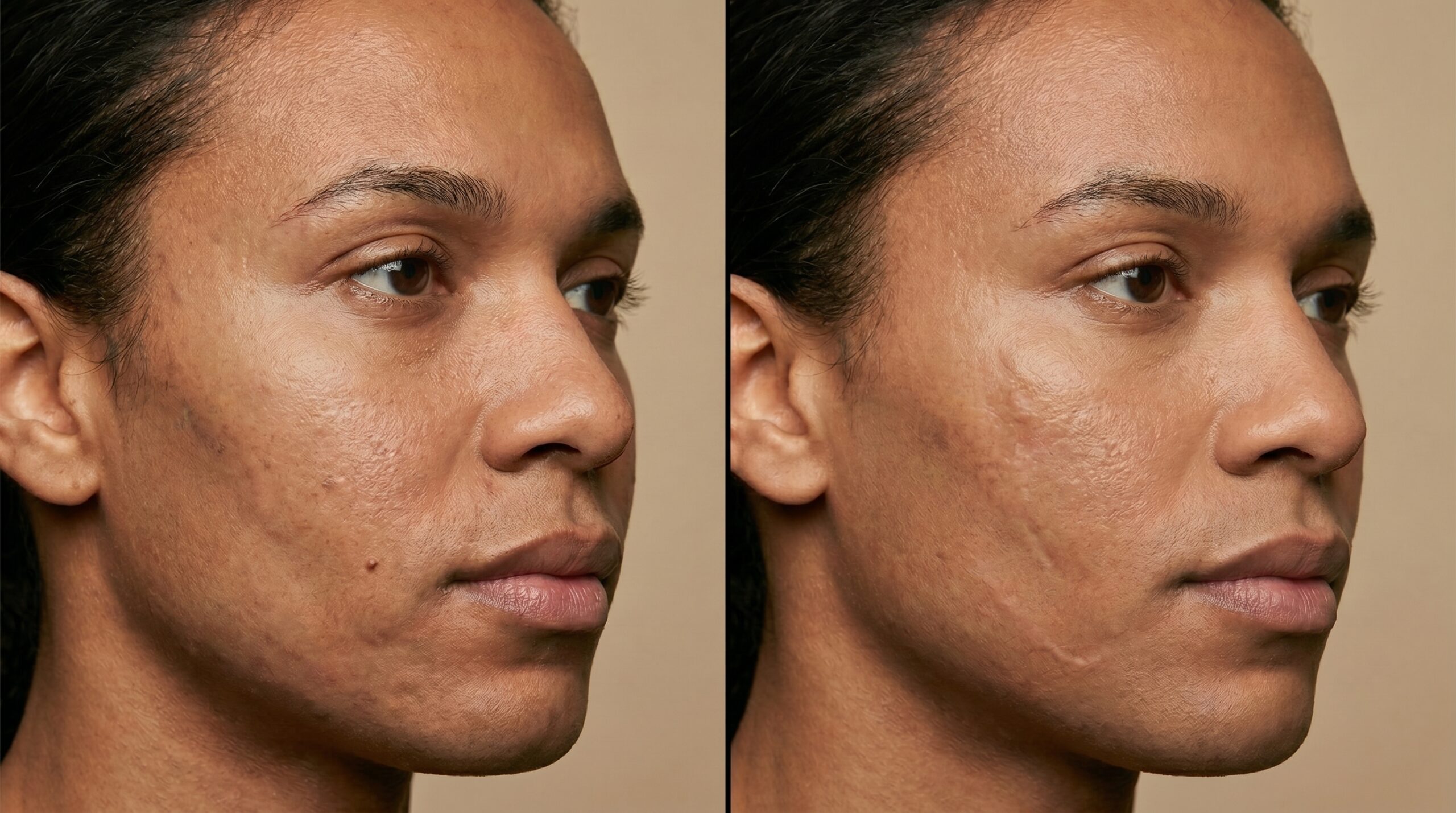
Individual results vary. Shave removal sites typically fade over 4 to 12 weeks; excision scars continue to mature and flatten over several months.
Every Mole Deserves
a Trained Eye.
When a mole changes, bleeds, or simply bothers you, the right response is a proper clinical evaluation — not reassurance from a photograph or a friend. At Couture Dermatology and Laser, Dr. Chinonso examines each lesion with dermoscopy and gives you a clear, evidence-based answer. Suspicious moles are excised and sent to pathology. Nothing is guessed.
Sat · By Appointment Only
"I came in with a mole that had been changing for about two months. Dr. Chinonso looked at it with a dermoscope, explained exactly what she was seeing, and removed it the same day. Pathology came back as a mildly dysplastic nevus with clear margins. The whole experience — from exam to results — was exactly what a dermatology appointment should be."
Marcus T.
Verified Patient · Beverly Hills
Frequently
Asked Questions
Direct answers to the questions patients most commonly ask before a mole check — on evaluation, removal methods, pathology, and what to expect from results.
No. Most moles are benign and require only monitoring. Removal is appropriate when a mole shows atypical dermoscopic or clinical features that raise concern, when it is a large congenital nevus with elevated lifetime risk, or when it is causing mechanical irritation or is cosmetically bothersome. A board-certified dermatologist will examine the lesion and recommend the appropriate course.
No, and this is a critical safety point. Any mole that looks irregular, has changed, or raises clinical concern must be excised with a scalpel and sent to pathology. Laser and cautery destroy the tissue needed for histological diagnosis. If a mole is suspicious, burning or lasering it can mask a melanoma and delay a life-saving diagnosis. At Couture Dermatology and Laser, concerning moles are always excised and sent to pathology.
ABCDE is the standard clinical screening tool: Asymmetry (one half doesn't match the other), Border (irregular, ragged, or poorly defined edges), Color (uneven distribution, multiple shades of brown, black, red, or white), Diameter (larger than 6mm, roughly the size of a pencil eraser), and Evolution (any change in size, shape, color, or a new symptom like bleeding or itching). The ugly-duckling sign — a mole that simply looks different from your other moles — is an equally important red flag.
Shave removal uses a thin blade to slice a raised, benign mole flush with the skin surface under local anesthesia. It leaves a small flat mark that fades over weeks to months. Surgical excision cuts the full thickness of skin around and beneath the lesion and is closed with sutures. Excision is used for flat moles, any lesion with atypical features, or any case where tissue needs to go to pathology for a complete histological margin assessment.
Most individual mole removals take 15 to 30 minutes including local anesthesia. Shave sites heal within 1 to 3 weeks and typically leave a small pink mark that continues to fade. Excision sites require suture removal at 7 to 14 days depending on location; a flat scar remains and its final appearance becomes apparent over several months. Patients should keep sites clean, avoid submerging them in water until healed, and apply SPF rigorously to the area.
Any mole that has changed in size, shape, or color, developed an irregular border, begun bleeding or itching, or simply looks different from your other moles warrants prompt evaluation. Annual full-skin examinations are recommended for anyone with more than 50 moles, a personal or family history of melanoma, fair skin, a history of significant sun exposure, or a prior history of atypical moles. When in doubt, have it checked — early detection of melanoma is the most important factor in outcome.