Basal Cell
Carcinoma
Basal cell carcinoma is the most common skin cancer — and also one of the most treatable when caught early. It grows slowly and rarely spreads, but it is locally destructive: left alone, it invades deeper tissue over time. At Couture Dermatology and Laser, diagnosis begins with a biopsy, not a guess, and treatment is selected precisely by subtype, location, and your individual anatomy.
primary BCC with Mohs
Dermatologist
most effective strategy

"A suspicious lesion that bleeds, scabs, and returns in the same spot deserves a biopsy — not watchful waiting."
Not All BCCs
Look the Same.
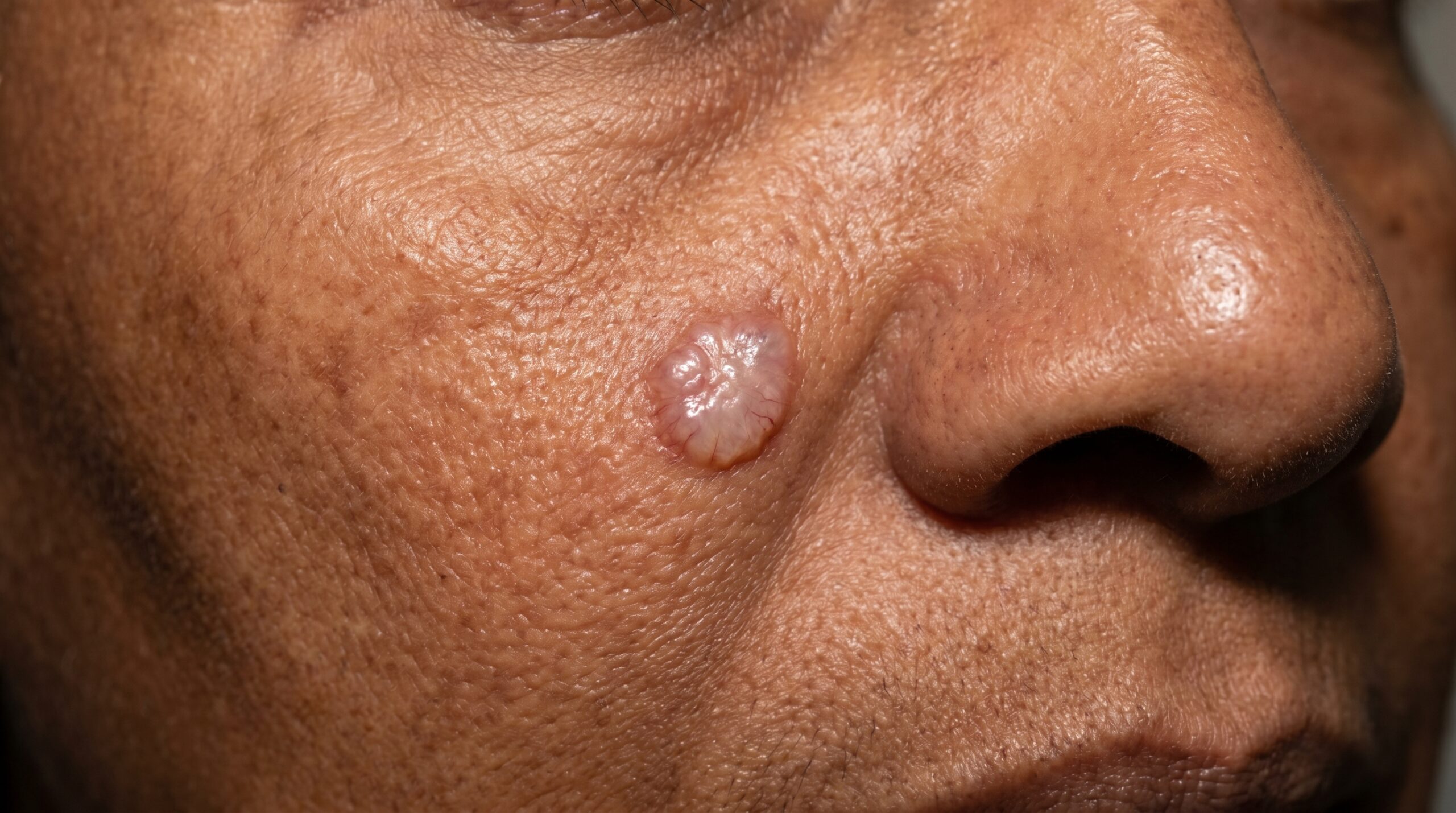
Basal cell carcinoma arises from the basal cells of the epidermis and presents in several distinct patterns. The classic nodular BCC appears as a pearly or translucent papule with rolled borders and fine surface blood vessels (telangiectasias) — often on the nose, cheek, or forehead. It may bleed with minimal trauma and crust over, only to return in the same spot.
Superficial BCC presents as a scaly, pink-red patch that can resemble eczema or psoriasis — often on the trunk or extremities. Morpheaform BCC is the most deceptive: it appears as a flat, scar-like, or ivory-white plaque with poorly defined edges. Because it infiltrates widely beneath the surface, it requires margin-controlled surgery and carries higher recurrence rates.
Regardless of presentation, the diagnostic pathway at Couture Dermatology and Laser is consistent: full skin exam, dermoscopy, and a definitive biopsy before any treatment decision is made.
From Diagnosis
to Clear Margins
Treating basal cell carcinoma correctly starts with understanding exactly what you are dealing with. Every step in this process is guided by pathology, not assumption.
Full Skin Examination
A thorough head-to-toe skin exam with dermoscopy evaluates all suspicious lesions in context. Sun-exposed areas receive particular attention, but a full-body check is standard at every visit.
Every new patient visitBiopsy & Subtype
A shave or punch biopsy provides definitive histologic diagnosis and identifies the specific subtype — nodular, superficial, or morpheaform — which determines the appropriate surgical approach.
Pathology-confirmed diagnosisMargin-Controlled Removal
Treatment is matched to risk. Mohs micrographic surgery for facial and high-risk tumors, standard excision for lower-risk sites, and ED&C or topical therapy for selected superficial lesions.
Treatment matched to subtypeRepair & Surveillance
Wound closure, reconstruction if needed, and post-operative scar care are addressed. A scheduled surveillance program follows — because one BCC is a meaningful predictor of future skin cancers.
6–12 month follow-up cycleThe Right Removal
for Every Tumor
No single removal technique is appropriate for all BCCs. Treatment selection depends on subtype, anatomic location, tumor size, and whether the lesion is primary or recurrent.
High-Risk & Facial Tumors
Mohs Micrographic Surgery
Mohs surgery removes the tumor one thin layer at a time. Each layer is mapped and examined under the microscope before proceeding — meaning 100% of the surgical margin is checked, not just representative sections. This produces the highest documented cure rates (approximately 99% for primary BCC) while sparing the maximum amount of normal tissue for reconstruction.
Lower-Risk & Truncal Lesions
Surgical Excision
Standard elliptical excision with clinically appropriate margins is an effective treatment for many BCCs on the trunk, extremities, and lower-risk facial sites. Pathology confirms margins after removal. Appropriate closure is planned at the same time to optimize the cosmetic outcome.
Small, Superficial, Low-Risk
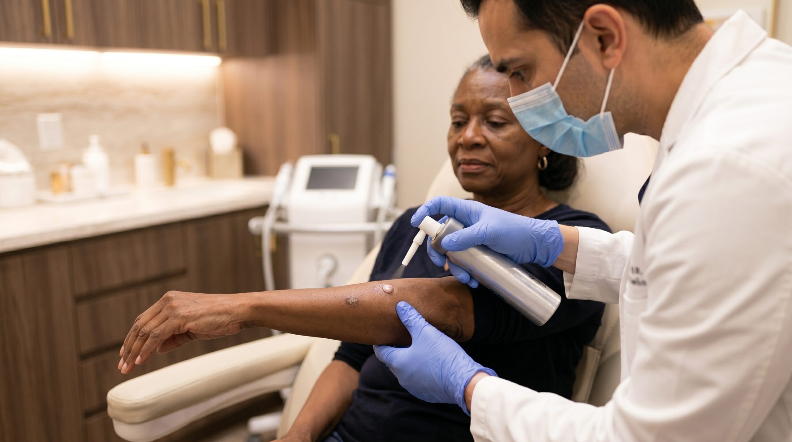
Electrodesiccation & Curettage
ED&C uses a curette to scrape away the tumor followed by electrodesiccation to destroy any remaining abnormal cells. It is appropriate for small, well-defined, superficial BCCs in low-risk anatomic locations — typically on the trunk or extremities away from critical structures.
Superficial Subtype, Selected Cases
Topical Therapy & PDT
Imiquimod cream and topical 5-fluorouracil (5-FU) activate an immune response or interfere with cell replication in superficial BCC. Photodynamic therapy (PDT) is an additional option in appropriate patients. These approaches are reserved for confirmed superficial subtype in patients where surgery is not preferred — they are not interchangeable with surgical removal for nodular or morpheaform disease.
Treat Early.
Protect What Matters.
Complete Margin Clearance
The primary goal of any BCC treatment is confirmed removal of the entire tumor. Mohs surgery achieves this by examining 100% of the margin in real time — so closure happens only once clear margins are confirmed under the microscope.
Tissue Preservation on the Face
Mohs surgery is specifically designed to remove the minimum amount of normal tissue necessary to achieve clear margins. On the nose, eyelids, lips, and ears — where tissue is limited — this distinction is clinically significant for both function and appearance.
Preventing Local Invasion
While BCC rarely metastasizes, an untreated or inadequately treated lesion continues to grow locally. Over years, this can mean invasion into cartilage, bone, or nerve structures — outcomes that are substantially more complex to manage than a straightforward early-stage removal.
Scar Management & Reconstruction
Removal of a BCC creates a wound that needs thoughtful closure. Dr. Chinonso discusses reconstruction options — primary closure, local flap, or skin graft — at your consultation, and post-operative scar treatments are available as part of your follow-up care.
Ongoing Skin Surveillance
A history of BCC meaningfully increases the likelihood of developing additional skin cancers — both BCC and squamous cell carcinoma. Regular full-body skin checks at Couture Dermatology and Laser allow new lesions to be identified and addressed at the earliest, most treatable stage.
"The lesion that bleeds and heals and bleeds again deserves a biopsy this week, not a photograph and a follow-up in six months."— Couture Dermatology and Laser
Mohs — primary BCC
Board-Certified
All skin cancer diagnosis and management is led by Dr. Chinonso Kagha Abisogun, MD, FAAD — a Fellow of the American Academy of Dermatology
Biopsy Before Treatment
No BCC is treated on clinical appearance alone — every lesion is confirmed by histopathology before a treatment plan is established
Subtype-Matched Care
Treatment selection — Mohs, excision, ED&C, or topical — is matched to the specific BCC subtype, location, and recurrence risk, not a default protocol
Structured Surveillance
Following treatment, a formal skin surveillance schedule is established — because one BCC is a known risk factor for additional skin cancers
How Basal Cell
Carcinoma Appears
BCC can look quite different depending on its subtype and anatomic location. These presentations are commonly seen in clinical practice — none should be dismissed without a proper skin exam.
Pearly Papule
A shiny, translucent bump with rolled edges and visible fine blood vessels on the surface — the classic nodular BCC appearance, most often on the face.
Non-Healing Sore
A lesion that bleeds, forms a crust, partially heals, then bleeds again in the same location. This recurrent cycle is a hallmark presentation of nodular BCC.
Scaly Pink Patch
A flat, red-pink, scaling lesion — superficial BCC — often found on the trunk, shoulders, or back. Frequently mistaken for eczema or a persistent irritation.
Scar-Like Plaque
A waxy, flat, or depressed area that resembles scar tissue without any prior injury — a characteristic of morpheaform BCC, which infiltrates broadly beneath the surface.
Pigmented BCC
A nodular BCC containing melanin pigment — appearing brown, blue, or black. Can be mistaken for a melanoma or seborrheic keratosis, underscoring the importance of dermoscopy and biopsy.
Recurrent BCC
A BCC returning at or near a previously treated site. Recurrent tumors often have subclinical extensions beyond the visible scar and require Mohs surgery for adequate margin control.
Is This Lesion
Worth Checking?
- Anyone with a skin lesion that bleeds spontaneously or with minimal friction, crusts over, and returns in the same location — regardless of how small it appears.
- Patients with fair skin, light eyes, a history of significant sun exposure or sunburns, or those who spent childhood in high-UV environments.
- Individuals with a personal or family history of skin cancer, or those with multiple actinic keratoses — known precancerous lesions that signal cumulative UV damage.
- Immunosuppressed patients, including organ transplant recipients, who face substantially elevated skin cancer risk and may develop multiple or aggressive lesions.
- Patients with a previously treated BCC who have not yet established a regular surveillance schedule — the risk of a second skin cancer is meaningfully elevated.
- Anyone who has noticed a new, changing, or unusual skin lesion and wants a definitive clinical evaluation, not a wait-and-see approach.
What to Expect at Consultation
A new patient with a suspicious lesion will receive a full skin examination with dermoscopy. If a biopsy is clinically indicated, it is typically performed at the same visit. Once pathology returns, Dr. Chinonso reviews the result with you directly, explains the subtype and its implications, and discusses the treatment options that are appropriate for your specific tumor.
No treatment is performed before you understand the diagnosis and have agreed on a plan. If Mohs surgery is indicated and requires a referral or scheduling at a specific surgical suite, that coordination will be managed for you.
Part of a Broader
Skin Health Plan
Basal cell carcinoma does not occur in isolation. The same UV damage that produced one BCC often signals wider changes in the skin that deserve attention alongside — and after — your primary treatment.
Squamous Cell Carcinoma
The second most common skin cancer, SCC can arise on actinic keratoses and carries a higher metastatic potential than BCC. Patients with one type of skin cancer are at elevated risk for the other.
Explore SCC treatment →Actinic Keratosis
Rough, scaly patches caused by UV damage that can progress to squamous cell carcinoma. Treating AKs reduces future skin cancer burden and is an important part of a proactive surveillance program.
Explore AK treatment →Melanoma Screening
A history of UV-induced BCC warrants careful evaluation of pigmented lesions at every surveillance visit. Full-body dermoscopy allows Dr. Chinonso to identify atypical moles and melanoma at the earliest stage.
Explore melanoma screening →Mole Evaluation
New, changing, or atypical moles require professional evaluation. Dermoscopy at Couture Dermatology and Laser provides far more diagnostic information than visual inspection alone — reducing unnecessary biopsies and ensuring none are missed.
Explore mole evaluation →Diagnosis to Healed Skin
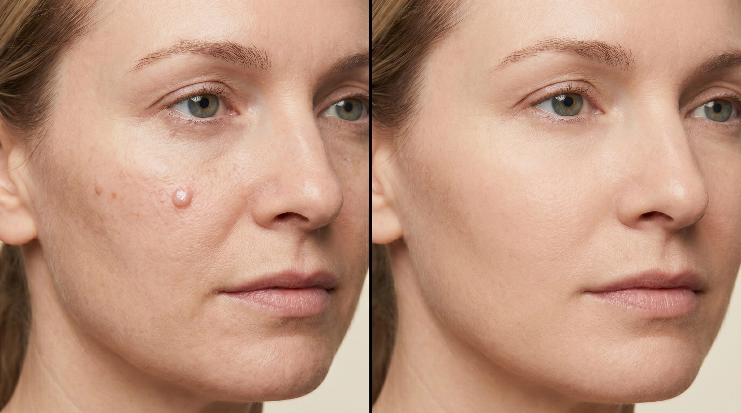
Individual results vary. Wound healing and final scar appearance depend on tumor size, location, closure technique, and patient-specific factors. Post-operative scar management is available as part of your care plan.
Diagnosing First.
Treating with Precision.
A suspicious lesion on the face or scalp does not get better by waiting. At Couture Dermatology and Laser, Dr. Chinonso performs a full skin examination and dermoscopy at your first visit, takes a biopsy if clinically indicated, and reviews the pathology with you before any surgical plan is discussed. You will understand your diagnosis completely before any procedure is scheduled.
Sat · By Appointment Only
"I had a small spot on my nose that my previous doctor kept watching. Dr. Chinonso biopsied it at my first visit — it was BCC. The Mohs procedure was far less daunting than I expected, and the repair looks better than I'd hoped. I just wish I hadn't waited so long."
Robert K.
Verified Patient · Beverly Hills
Frequently
Asked Questions
Direct answers to the questions patients ask most often when they come in with a suspicious skin lesion — on diagnosis, treatment options, what surgery involves, and what happens after.
Diagnosis requires a skin biopsy — there is no reliable way to confirm BCC by appearance alone. At your consultation, Dr. Chinonso performs a full skin exam and dermoscopy to evaluate suspicious lesions, then takes a shave or punch biopsy for pathologic confirmation. The biopsy report also identifies the subtype (nodular, superficial, morpheaform), which directly determines which treatment is appropriate.
Mohs micrographic surgery removes the tumor layer by layer while the surgeon examines 100% of the margins under a microscope in real time. This approach achieves the highest cure rates for primary BCC — approximately 99% for primary tumors — and is the standard of care for BCCs on the face, ears, nose, and scalp; for recurrent tumors; for morpheaform or infiltrative subtypes; and for lesions in cosmetically sensitive or functionally critical areas.
Basal cell carcinoma grows slowly and rarely metastasizes — the lifetime metastasis risk is less than 0.1% for typical cases. However, it is locally destructive. An untreated or recurrent BCC can invade deeply into bone, cartilage, and nerves over time, particularly on the face. Early detection and complete removal prevent this and are the primary reasons not to delay treatment of a suspicious lesion.
Treatment is matched to tumor subtype, location, and patient factors. Standard surgical excision with adequate margins is appropriate for many lower-risk lesions on the trunk and extremities. Electrodesiccation and curettage (ED&C) is an option for small, superficial BCCs in low-risk locations. Topical imiquimod or 5-fluorouracil (5-FU) can treat superficial BCCs in selected patients. Radiation therapy is an alternative for patients who cannot tolerate surgery. Hedgehog-pathway inhibitors (vismodegib, sonidegib) are reserved for advanced or locally extensive disease.
Any skin cancer removal leaves some degree of scarring — the extent depends on the tumor's size and location, the surgical technique, and how the wound is closed. Mohs surgery is designed to remove the minimum necessary tissue while achieving clear margins, which preserves normal tissue for repair. Dr. Chinonso discusses reconstruction and scar management options at your consultation, including primary closure, flaps, and post-operative scar treatments as appropriate.
A history of BCC significantly increases the risk of developing additional skin cancers. We recommend full-body skin surveillance every 6 to 12 months, depending on your skin type, prior history, and sun exposure. Patients with multiple prior BCCs, immunosuppression, or a history of significant UV exposure may be seen more frequently. Ongoing surveillance is as important as the initial treatment.