Squamous Cell
Carcinoma
Squamous cell carcinoma is the second most common skin cancer. Caught early — before it has any opportunity to deepen or spread — the prognosis is excellent and the treatment straightforward. At Couture Dermatology and Laser, every suspicious lesion receives a thorough evaluation, biopsy confirmation, and a removal strategy matched precisely to its size, location, and risk profile.
skin cancer
early-stage SCC
Dermatologist

"A rough, persistent patch that won't heal deserves a biopsy — not watchful waiting."
What Is Squamous
Cell Carcinoma?
Squamous cell carcinoma arises from keratinocytes — the cells that make up the outer layers of skin. Cumulative ultraviolet exposure is the dominant driver, which is why SCC appears most often on chronically sun-exposed areas: the face, scalp, ears, lips, back of the hands, and forearms. Many cases develop from actinic keratoses, the rough, scaly precancerous patches that signal significant UV damage to the underlying skin.
Unlike basal cell carcinoma, squamous cell carcinoma carries a higher — though still generally low — potential to spread to regional lymph nodes and beyond. That risk is not uniform: small, low-risk SCCs on the trunk behave quite differently from large or recurrent tumors on the lip or ear, and from any SCC in a patient on long-term immunosuppression (such as organ transplant recipients, who face a markedly elevated SCC burden). Location, size, depth, histologic features, and immune status all factor into how aggressively a given tumor is managed.
The earliest form, SCC in situ (Bowen's disease), is confined entirely to the epidermis and has not yet invaded deeper tissue. It is treatable with less invasive methods when caught at this stage — underscoring why annual full-body skin examinations and prompt evaluation of suspicious lesions matter.
From Evaluation
to Clear Margins
Squamous cell carcinoma treatment follows a deliberate sequence — diagnosis first, removal second, reconstruction and surveillance third. No step is skipped.
Full-Body Skin Exam
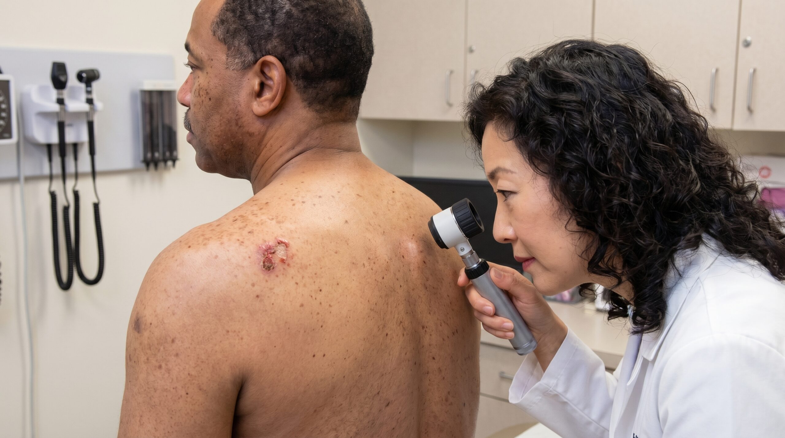
A thorough head-to-toe examination with dermoscopy identifies the primary lesion, screens for additional suspicious areas, and assesses regional lymph nodes clinically where indicated.
Dermoscopy includedBiopsy & Pathology
A skin biopsy is performed and sent for histopathologic analysis. Treatment planning waits for confirmed pathology — including tumor subtype, depth, and any high-risk histologic features.
No treatment without diagnosisMargin-Controlled Removal
Treatment is selected based on tumor size, location, and risk. Mohs micrographic surgery, standard excision, ED&C, or topical/PDT for in situ disease — each chosen for its appropriateness, not convenience.
Treatment matched to riskReconstruction & Surveillance
Wound closure, scar management, and ongoing surveillance skin exams every 3 to 6 months for high-risk patients. Sun protection counseling at every visit.
Lifelong monitoringRemoval Strategies
by Risk & Location
The best treatment for squamous cell carcinoma depends on where the tumor is, how large it is, and what its pathology shows. These four approaches cover the spectrum of clinical need.
High-Risk & Facial Tumors
Mohs Micrographic Surgery
Mohs surgery removes the tumor in precisely mapped layers, and 100% of the surgical margins are examined microscopically before closure. This staged process continues until no residual cancer cells remain at the edges. It achieves the highest cure rates available for SCC and is preferred for tumors on the face, scalp, ears, and lips — where both complete removal and tissue conservation matter — as well as for recurrent, large, or histologically aggressive tumors.
Low-to-Moderate Risk Lesions
Standard Surgical Excision
Excision with clinically appropriate margins — typically 4 to 6 mm for low-risk SCC — followed by standard pathologic margin evaluation. Suitable for well-defined tumors on low-risk sites. Reconstruction is performed at the time of excision when margins are assessed as adequate, or as a staged procedure when needed.
SCC In Situ / Small Low-Risk Lesions
ED&C, Topical & PDT
Electrodesiccation and curettage (ED&C) is appropriate for small, low-risk lesions on non-critical skin sites in appropriate patients. SCC in situ (Bowen's disease) may also be treated with topical 5-fluorouracil or imiquimod, or with photodynamic therapy — options selected based on lesion size, patient health, and location.
Non-Surgical Candidates & Advanced Disease
Radiation & Oncology Coordination
Radiation therapy is an appropriate alternative for patients who cannot undergo surgery or where surgery would cause significant functional impairment. High-risk or locally advanced SCC requiring lymph node evaluation, sentinel lymph node biopsy, or systemic immunotherapy (cemiplimab) is coordinated with oncology specialists — ensuring a complete care pathway.
Caught Early.
Treated Completely.
High Cure Rates for Early-Stage SCC
Squamous cell carcinoma diagnosed and removed at an early, localized stage carries excellent cure rates. The clinical imperative is to find it before it has the opportunity to grow deeper or reach the lymph nodes, where management becomes considerably more complex.
Complete Margin Clearance
The primary goal of surgical treatment is removal with pathologically clear margins. Incomplete excision — leaving residual tumor cells at the wound edge — is the main driver of local recurrence. Mohs surgery eliminates this uncertainty for high-risk tumors by confirming margins intraoperatively, layer by layer.
Tissue-Sparing Reconstruction
On the face, ears, nose, and lips, the tissue conservation inherent to Mohs surgery allows reconstruction that restores form and function with minimal distortion. Closure options — primary repair, flap, or graft — are selected for each anatomic site after the tumor is confirmed clear.
Appropriate Risk Stratification
Not all SCCs carry the same risk. Organ transplant recipients, patients on chronic immunosuppression, and those with large, recurrent, or poorly differentiated tumors require a more vigilant management approach. Identifying high-risk features at the outset allows the treatment plan — and the surveillance schedule — to match the actual risk.
Long-Term Skin Cancer Surveillance
A history of squamous cell carcinoma is itself a risk factor for subsequent skin cancers — both SCC and other types. Regular full-body skin exams, patient education on warning signs, and strict sun protection reduce the likelihood of missing a new lesion at its most treatable stage.
"The best outcome in squamous cell carcinoma is not dramatic — it is a clean pathology report and a patient who keeps their surveillance appointments."— Couture Dermatology and Laser
with Mohs surgery
Board-Certified
Every squamous cell carcinoma case is evaluated personally by Dr. Chinonso Kagha Abisogun, MD, FAAD — a Fellow of the American Academy of Dermatology
Biopsy First
No lesion is treated without histopathologic confirmation. Dermoscopy narrows the differential, but pathology makes the diagnosis
Risk-Matched Care
Treatment intensity reflects the actual risk profile of each tumor — from minimal procedures for SCC in situ to Mohs and oncology coordination for high-risk disease
Lifelong Surveillance
Skin cancer treatment does not end at clear margins — structured follow-up keeps future lesions from reaching an advanced stage
Common Sites &
Presentations
Squamous cell carcinoma favors areas of chronic sun exposure and has several characteristic presentations. Knowing what to look for — and where — is the first step toward catching it early.
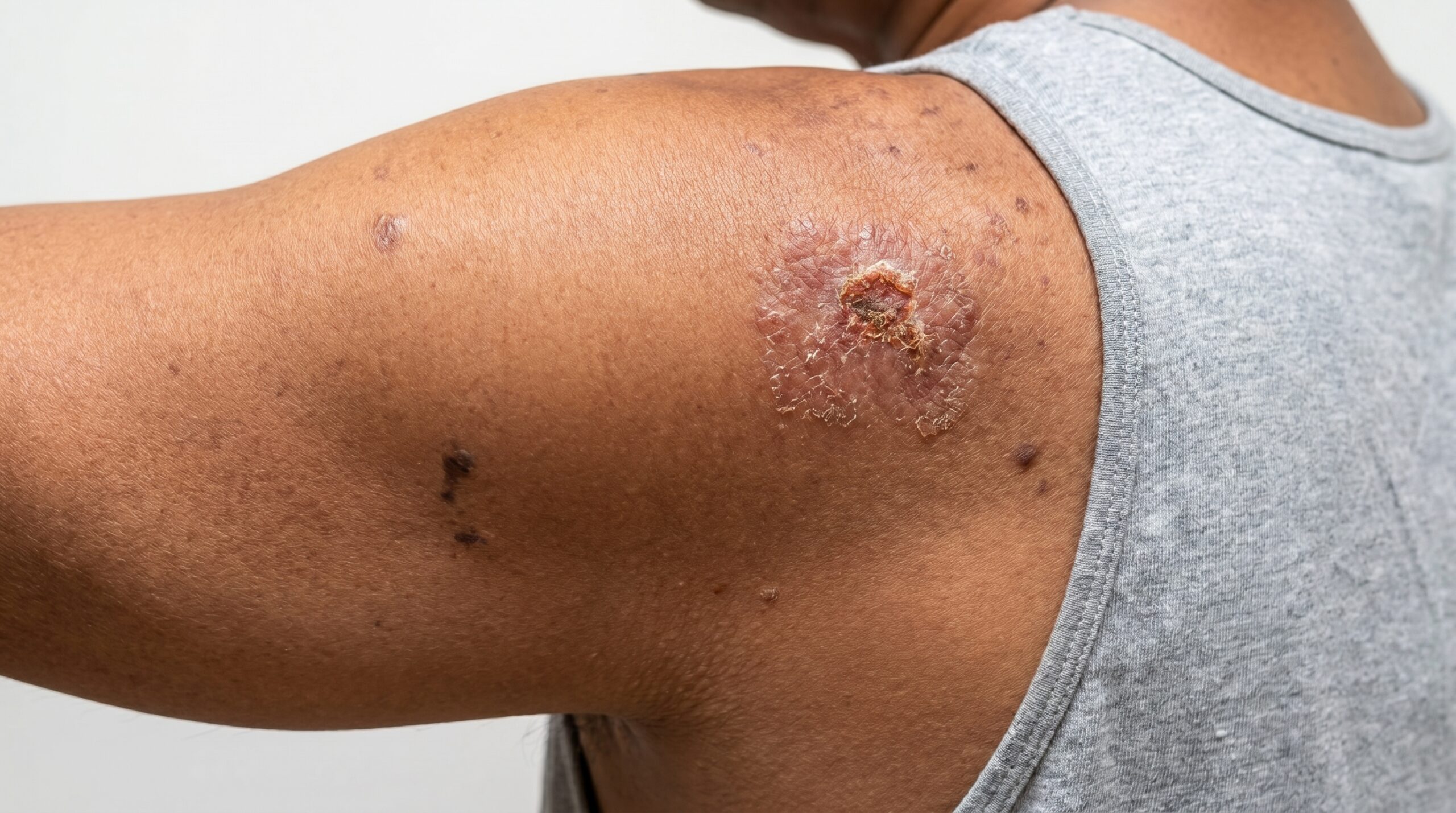
Scaly or Crusted Patch
A rough, keratotic plaque that may bleed with minor trauma. Often arises on a background of actinic keratoses. The most common early presentation.
Firm Red Nodule
A raised, indurated lesion — sometimes tender — on sun-exposed skin. May ulcerate centrally. Often seen on the scalp, face, or hands in older patients.
Non-Healing Wound
A sore that bleeds, scabs, and refuses to close over several weeks. Any wound that does not heal within a month warrants dermatologic evaluation.
Lip & Ear Tumors
Higher-risk locations where SCC has greater metastatic potential. Any persistent lesion on the vermilion lip or pinna of the ear should be biopsied promptly.
SCC In Situ (Bowen's Disease)
A flat, slowly enlarging red-brown scaly plaque confined to the epidermis. The earliest and most treatable form — managed with less invasive approaches when identified at this stage.
Immunosuppressed Patients
Organ transplant recipients and others on long-term immunosuppression develop SCC at dramatically higher rates and with more aggressive behavior. Dedicated skin cancer surveillance programs are essential for this population.
Who Should Be
Seen Promptly?
- Anyone with a rough, scaly, or crusted skin lesion that has persisted for more than a few weeks — particularly on sun-exposed areas.
- Patients with a known history of actinic keratoses or a prior skin cancer of any type, as their cumulative UV damage places them at higher baseline risk.
- Organ transplant recipients and patients on chronic immunosuppressive therapy, who should be on a structured skin cancer surveillance program rather than annual exams alone.
- Those who notice a wound that bleeds easily, scabs repeatedly, or has not healed within four weeks despite basic wound care.
- Fair-skinned individuals with significant lifetime sun exposure, outdoor occupational history, or a history of tanning bed use.
- Patients who have previously been told they have a "precancerous" lesion and want a clear plan for monitoring or treating it before it progresses.
What to Expect at Your Evaluation
An evaluation for squamous cell carcinoma involves a thorough full-body skin examination — not just the area of concern. Dermoscopy allows close inspection of lesion structure before any decision is made. If a biopsy is indicated, it is performed at the same visit when appropriate, and results are typically available within one to two weeks.
Treatment planning is discussed only after pathology is confirmed. Dr. Chinonso will review the biopsy findings, explain the options suited to your specific tumor, and give you a direct, honest assessment of what the treatment involves, what the expected outcome is, and what surveillance will look like going forward.
Related Conditions
We Also Address
Squamous cell carcinoma rarely exists in isolation. Patients with SCC often have additional skin cancers, precancerous lesions, or pigmented concerns that benefit from coordinated evaluation and treatment.
Basal Cell Carcinoma
The most common skin cancer, also driven by UV exposure and frequently diagnosed alongside SCC in the same patient. Managed with similar margin-controlled techniques, including Mohs surgery for high-risk lesions.
Learn about BCC treatment →Melanoma
The most serious form of skin cancer. Patients with a history of SCC benefit from comprehensive pigmented lesion evaluation — dermoscopy and biopsy when indicated — to ensure concurrent melanoma is not missed.
Learn about melanoma →Actinic Keratosis
The most direct precursor to squamous cell carcinoma. Treating actinic keratoses reduces the field-cancerization burden and lowers the probability of progression to invasive SCC. Multiple treatment modalities are available.
Learn about actinic keratosis →Moles & Pigmented Lesions
A thorough skin examination for SCC naturally includes full evaluation of moles and pigmented lesions. Dermoscopy distinguishes benign nevi from dysplastic or melanocytic lesions requiring biopsy.
Learn about mole evaluation →Diagnosis to Healed Skin
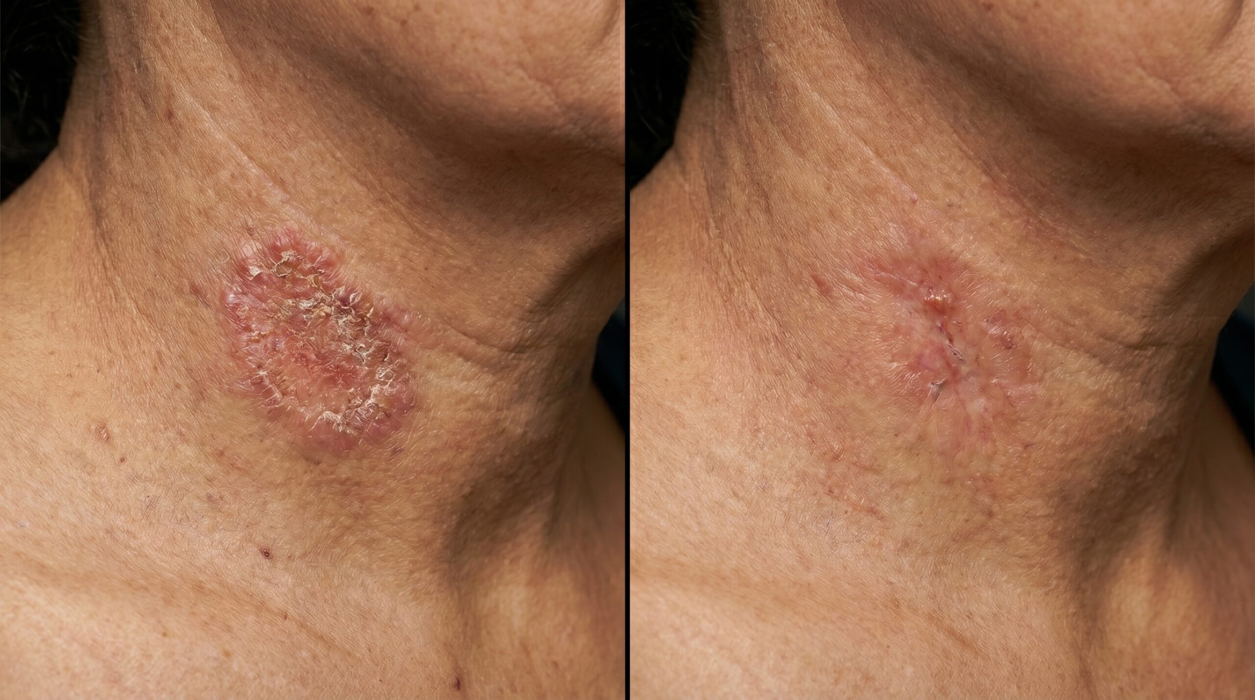
Individual outcomes vary based on tumor size, location, and patient healing. Results shown are following confirmed clear-margin removal and standard wound healing. Photography is for educational purposes and does not constitute a treatment guarantee.
A Lesion That Concerns You
Deserves an Answer.
Skin cancer is manageable when it is found early. At Couture Dermatology and Laser, Dr. Chinonso evaluates every suspicious lesion with dermoscopy, biopsies when indicated, and builds a treatment plan grounded in pathology — not guesswork. No diagnosis is given without proper confirmation, and no procedure is recommended without a direct conversation about its purpose and expected outcome.
Sat · By Appointment Only
"I noticed a spot on my temple that wasn't healing. Dr. Chinonso biopsied it, confirmed it was squamous cell carcinoma, and walked me through every option clearly and calmly. The Mohs procedure was done in the office, the margins were clear, and the scar is minimal. I'm grateful I didn't wait."
Richard T.
Verified Patient · Beverly Hills
Frequently
Asked Questions
Direct answers to what patients most often ask before their squamous cell carcinoma evaluation — on diagnosis, surgery, risk, and what comes after.
Diagnosis requires a skin biopsy. A board-certified dermatologist will perform a full skin examination and may use dermoscopy to identify suspicious lesions before taking a small tissue sample for pathology. No treatment is performed without biopsy confirmation.
SCC in situ, also called Bowen's disease, is the earliest form of squamous cell carcinoma. The abnormal keratinocytes are confined to the upper layer of the skin and have not yet invaded deeper tissue. It carries a lower risk of spread than invasive SCC and may be treated with topical therapy, photodynamic therapy, or a minor procedure rather than surgery.
Higher-risk features include tumors larger than 2 cm, tumors on the lip or ear, poorly differentiated histology, perineural invasion, deep invasion, and a history of previous SCC in the same area. Immunosuppressed patients — particularly organ transplant recipients — face a substantially elevated risk of both development and spread. These cases typically warrant a more aggressive treatment approach and closer surveillance.
Mohs micrographic surgery is a specialized technique in which the surgeon removes the tumor in thin layers, immediately examining 100% of the surgical margins under a microscope before proceeding. This continues until no cancer cells remain at the edges. It is preferred for SCCs on the face, scalp, ears, and lips — where tissue conservation matters — and for recurrent, large, or histologically aggressive tumors. Cure rates for SCC treated with Mohs are among the highest available.
After excision, wound care instructions are provided, and sutures are typically removed within 7 to 14 days. Scar management begins once the wound is fully healed. Pathology confirms clear margins. Ongoing surveillance is essential: patients with a history of SCC are at higher risk for additional skin cancers and are scheduled for full-body skin checks every 3 to 6 months depending on individual risk.
Complete removal with clear margins is the goal of initial treatment and significantly reduces the risk of local recurrence. However, patients with a history of SCC remain at elevated lifetime risk for developing new skin cancers due to the same underlying UV damage and immune factors. Strict daily sun protection — broad-spectrum SPF 30 or higher, protective clothing, and hat use — combined with regular dermatology surveillance gives patients the best chance of catching any new lesions at the earliest stage.